
A team of scientists led by the Department of Applied Physics at Osaka University, the Department of Physics and Electronics at Osaka Prefecture University, and the Department of Materials Chemistry at Nagoya University used photoinduced force microscopy to map out the forces acting on quantum dots in three dimensions. By eliminating sources of noise, the team was able to achieve subnanometer precision for the first time ever, which may lead to new advances in photocatalysts and optical tweezers.
Force fields are not the invisible barriers of science fiction but are a set of vectors indicating the magnitude and direction of forces acting in a region of space. Nanotechnology, which involves making and manipulating tiny devices such as quantum dots, sometimes uses lasers to optically trap and move these objects. However, the ability to analyze and handle such small systems requires a better way to visualize the 3D forces acting on them.
Now, a team of researchers at Osaka University, Osaka Prefecture University, and Nagoya University has shown for the first time how photoinduced force microscopy can be used to obtain 3D force field diagrams with subnanometer resolution. "We succeeded in imaging the optical near-field of nanoparticles using a photoinduced force microscope. This measures the optical force between the sample and the probe caused by light irradiation," first author Junsuke Yamanishi says.
Laser light was directed on a quantum dot placed underneath an atomic force microscopy tip. Moving the dot relative to the tip allowed the microscope to map out the 3D photoinduced force field. The team was able to achieve such a high level of precision using a few experimental improvements. They used ultra-vacuum conditions to increase the force sensitivity, and employed heterodyne frequency modulation, which involves mixing two other frequencies, to greatly reduce the impact of thermal heating. "We reduced the photothermal effect with this unique technology and achieved a resolution of less than one nanometer for the first time ever," senior author Yasuhiro Sugawara says.
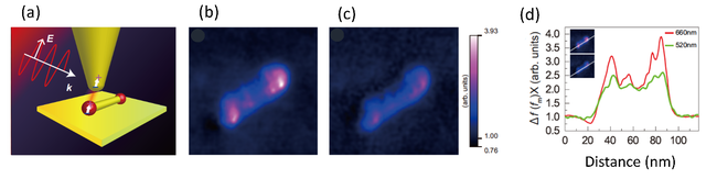
Fig.1 (a) Schematic image of photoinduced force microscopy. (b)(c) Photo-induced force microscopy images of a quantum dot measured using different wavelengths (600 nm,520 nm). (d) Photoinduced force profiles for the images. This reflects the electronic energy structure designed for photocatalysis. (credit: Osaka University)
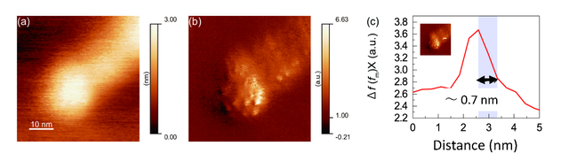
Fig.2 (a) Atomic force microscopy image of a quantum dot. (b) Photoinduced force microscopy image at 660 nm. (c) Photoinduced force profiles for the image. A spatial resolution of less than 1 nm has been obtained. (credit: Osaka University)
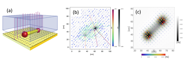
Fig. 3 (a) 3D force field mapping of the photoinduced force. (b) Experimentally obtained 3D photoinduced force field map using the laser with 660 nm wavelength. Colored arrows indicate the magnitude and direction of the force in the plane. The black and white shading indicates the magnitude of the force in the height direction. (c) Theoretically calculated 3D photoinduced force field map. The tendency to explain the results of experiments well is evident. (credit: Osaka University)
This research may represent a fundamentally new technology for the design and evaluation of functional nanomaterials. It can also help supplement the toolbox of methods available to scientists working with photocatalysts and optical functional devices to move them using lasers.
Read the original article on Nagoya University.