Our bodies have evolved formidable barriers to protect themselves against foreign substances – from our skin, to our cells and every component within the cells, each part of our bodies has protective layers. These defences, while essential, pose a significant challenge for pharmaceutical drugs and therapies, such as vaccines, that have to bypass multiple barriers to reach their targets.
Although these barriers are vitally important in pharmaceutical science and drug design, much is still unknown about them and how to overcome them.
In a recent study, researchers from Xi’an Jiaotong-Liverpool University and Nanjing University in China, and Western Washington and Emory University in the US, shed some light on why the delivery of therapeutics to cells can be so difficult.
Overcoming barriers
With Covid-19 vaccines, which hundreds of millions of us have been injected with, mRNA has to be enclosed within protective fatty bubbles – lipid nanoparticles – so it can pass through the body’s defences and reach the intended target in our cells.
Some types of cells, such as stem cells, immune cells, and nerve cells, have barriers that are particularly difficult to overcome, so the delivery of particles into these cells is even more challenging.
In the study, published in the American Chemical Society’s journal ACS Nano, the researchers combined cutting-edge microscopy techniques to track the delivery of nanoparticles, which are often used for drug delivery, into stem cells in real-time.
Their findings suggest that, in certain types of cells, nanoparticles become ‘trapped’ within bubble-like vesicles and so are prevented from reaching their intended target.
The team used their findings to create a mathematical model that can predict how efficient the delivery of nanoparticles into cells will be, and aid the design of future therapies.
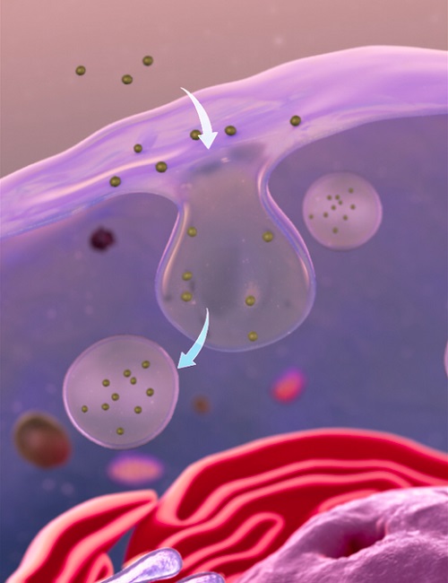
In order to gain entry to a cell, nanoparticles can be engulfed by the membrane surrounding the cell, forming bubble-like vesicles - endocytosis.
Dr Gang Ruan, a corresponding author of the study, says: “We have broken down the delivery process of particles into cells into individual steps, so we can visualise each step and create a window into the mechanisms used by these cells to protect themselves.
“To design improved delivery methods for therapies, we need a quantitative understanding of how parts of the cell and nanoparticles interact. Like a great bioengineer I knew once said, if you were to design an aeroplane, you’d have to analyse the aerodynamics of each part before building the plane.
“By finding the bottleneck in the delivery of nanoparticles into cells, our findings will pave the way for more targeted and innovative therapies that use tailored delivery, potentially for individual patients.”
Out for delivery
Previously, imaging of nanoparticle delivery in cells has been limited because of the required quick speed and small scale. However, the multidisciplinary team were able to use their different fields of expertise to create innovative ways to overcome these hurdles. They combined two types of microscopy analysis, previously only used separately, to enable them to study the entire delivery process.
Xuan Yang, who shares lead authorship of the study with Dr Xiaowei Wen, says: “We were able to track the movement of the nanoparticles on a pixel by pixel basis, in real-time, and therefore visualise the movement of the nanoparticles across membrane barriers and as they entered each compartment of the stem cells.”
Although the process of delivery of nanoparticles into these cells is complex and made up of several mechanisms, by visualising and then chemically modifying each step of the process, the team identified the critical stage that prevents delivery of the nanoparticles to their cell targets.
In order to gain entry to a cell, nanoparticles can be engulfed by the membrane surrounding the cell, forming bubble-like vesicles. In many cell types, the nanoparticles would escape from these bubbles once inside the cell. However, in some extra-protected cells, such as the stem cells used in this study, the nanoparticles seem to get trapped inside the vesicles and are unable to escape. This means they cannot enter the cell and reach their target.
The researchers combine their observations and analysis in a mathematical model that can predict how efficiently and quickly particles would go through each step of delivery and enter a cell.
“Our model can be used to predict what the concentration of the nanoparticles will be, at a particular location in the cell, at a particular time,” says Dr Wen.
“The general method of this model can be used to incorporate different types of nanoparticles and cells to better understand the delivery mechanisms used to pass into cells. For example, predicting how well lipid nanoparticles in the Covid-19 vaccines will deliver mRNA into a cell.”
Dr Steven Emory, who is also a corresponding author of the study, adds: “Being able to map out the different components and inner workings that make up the delivery pathways in real-time leads to understanding how to control these pathways. This could open up some really exciting things in terms of therapeutics.
“We hope our new tools and understanding have created an initial foothold for the system, from where we, and other researchers, can begin climbing and start exploring.”
Read the original article on Liverpool University.