A team of scientists, led by Xiling Shen, Ph.D., Chief Scientific Officer, and Professor at the Terasaki Institute for Biomedical Innovation (TIBI), has reached new levels in patient model development. They have developed improved methods for generating micro-organospheres (MOS) and have shown that these MOS have superior capabilities for a variety of clinical uses. As documented in a recent publication in Stem Cell Reports, their MOS can be used as patient avatars for studies involving direct viral infection, immune cell penetration and high-throughput therapeutic drug screening, something that is not obtainable with conventional patient-derived models.
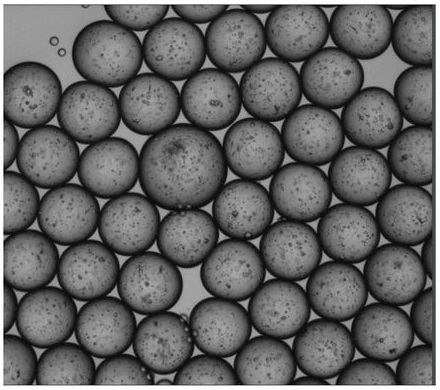
Picture depicting Micro-organospheres (MOS) encapsulating primary tissue derived cells prior to demulsification.
Dr. Shen’s team has developed emulsion microfluidic technology for creating MOS, tiny, nanoliter-sized basal membrane extract (BME) droplets composed of tissue cell mixtures which can be generated at a rapid pace from an automated device. After the droplets are created, excess oil is removed by an innovative membrane demulsification process, leaving behind thousands of viscous, uniformly sized droplets which contain tiny 3D tissue structures.
The team went on to demonstrate unique MOS capabilities and features in several first-of-its-kind experiments. They were able to show that the MOS could be created from a variety of different tissue sources and the resultant MOS had retention of histopathological morphology, capacity for differentiation and genetic expression, and the ability to be frozen and sub-cultured, as in conventional organoids.
Experiments were conducted to test the ability to infect MOS with viruses. Unlike with conventional organoids, MOS can be directly infected with viruses without the removal and suspension of cells from its surrounding BME scaffold, hence recapitulating the process of viral infection of the host tissue. Dr. Shen’s team was able to create a MOS atlas of human respiratory and digestive tissues from patient autopsies and infect them with SARS-COV-2 viruses, followed by drug screening to identify drugs that block viral infection and replication within those tissues.
MOS also provide a unique platform for studying and developing immune cell therapy. Within natural diffusion limit of vascularized tissue, tumor-derived MOS allowed sufficient penetration by therapeutic immune T-cells such as CAR-T, enabling a novel T cell potency assay to assess tumor killing by the engineered T-cells. Such a model would be highly useful in investigating tumor responsiveness and in developing anti-tumor immune cell therapies.
MOS could be further integrated with deep-learning imaging analysis for rapid drug testing of small and heterogeneous clinical tumor biopsies. Moreover, the algorithm was able to distinguish cytotoxic vs. cytostatic drug effects and drug-resistant clones that will give rise to later relapse. This groundbreaking capability will pave the way for MOS to be used in the clinic to inform therapeutic decisions.
Read the original article on Terasaki Institute for Biomedical Innovation (TIBI).