2022-11-16
Visited : 1316
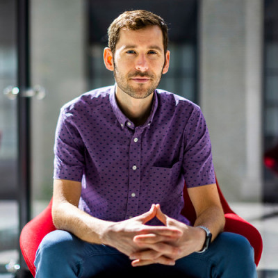
With new techniques in electron microscopy, James LeBeau explores the nanoscale landscape within materials to understand their properties.
To explain why he loves electron microscopy, Associate Professor James LeBeau uses an analogy: He likens the technique, which uses beams of electrons to illuminate materials at a scale thousands of times smaller than conventional microscopes, to the inverse of astronomy.
“It’s discovering things that no human has ever seen before that really captures the imagination,” LeBeau says. “There is a beauty to the way atoms are arranged in materials, particularly at defects, which give rise to all sorts of material behavior.”
LeBeau has used that passion to develop new techniques for collecting and interpreting data in electron microscopy that can be used to describe materials more comprehensively. He’s applied those techniques to explain materials’ behavior in fields from electronics and optics to energy storage, quantum computing, and more.
“Beyond explaining material properties, there’s also a significant computational component to electron microscopy as it’s used to analyze data that may have been overlooked previously and to make conclusions about the data in new ways. And, with the creation of the college of computing, it’s an exciting time to be at MIT,” he says.
Discovering a passion
LeBeau became interested in engineering while helping his father build and repair things around the house, and he discovered a love for science at a young age.
“Science can provide an explanation of the world around us beyond supernatural beliefs,” LeBeau says. “For me, science was about making sense of the world.”
LeBeau first learned about materials science through the technical high school he attended in Indiana. But it wasn’t until he was an undergraduate at Rensselaer Polytechnic Institute in New York that a few pivotal experiences helped set his course in life.
During his first year, he participated in a project using data science to predict material properties.
“After that I was hooked, and at that point I knew I wanted to go the academic route,” he recalls. “Just being able to explore things and have that academic freedom really appealed to me.”
A few years later, in 2005, LeBeau participated in a summer research program for undergraduates at what is now the Materials Research Laboratory at MIT. The experience, in which he integrated biopolymers into a casting process, stoked his interest in using materials science for sustainability. The passion of the researchers around MIT also left a lasting impression on him.
Finally, as a senior, LeBeau got his first taste of electron microscopy.
“We'd be in the lab in the middle of the night analyzing these materials, and that excitement caught my attention pretty early on,” LeBeau says. “It didn't really matter how much I was working — I loved doing it, and that set the stage for the rest of my career.”
During his PhD at the University of California at Santa Barbara, LeBeau was part of a team that showed that scanning transmission electron microscopy theory and experiment are in very good agreement and, in turn, that attograms (one millionth of a trillionth of a gram) of material could be weighed directly from electron microscopy images without the need for external microscope calibration standards.
LeBeau also discovered a passion for cycling through the mountains near UC Barbara’s campus, an activity he continues by biking thousands of miles a year, including to MIT nearly every day regardless of the weather.
After his PhD, LeBeau accepted a faculty position at North Carolina State University, where he worked for eight years before a similar position opened up at MIT in 2019.
Since his move to MIT, LeBeau has helped the Institute adopt state-of-the-art electron microscopy equipment that researchers from across campus have taken advantage of in MIT.nano and elsewhere.
“As an electron microscopist, the equipment I use is extremely expensive to maintain and necessitates that it becomes a shared resource. I’m happy that’s the case because ultimately users from across campus benefit from these tools and advance their science through this shared infrastructure,” LeBeau says. “More broadly, the microscope routinely challenges what people thought they knew about the materials they are studying. The results are always exciting.”
Creativity and quantification
When it’s his group’s turn on the microscope, LeBeau says they try to go after hard problems that require new ways of collecting and interpreting data.
“We choose questions that are not easy to answer through other methods and that require new ways to extract information from our datasets to make conclusions,” LeBeau says.
One type of material LeBeau has studied is relaxor ferroelectrics, which are used for applications including ultrasounds, actuators, and energy storage. The materials have been studied for decades but are extremely heterogeneous at the nanoscale, making it difficult to explain their electromechanical properties. By analyzing the materials’ structure using new electron microscopy techniques, LeBeau’s group was able to explain its properties in a way that could help create more sustainable versions of the material, which currently contain lead.
“Impact is always at the forefront of everything we do,” LeBeau explains. “When we go after problems, the application space is very important because it tells us if the insights can change the way an entire space operates.”
One area of LeBeau’s research explores ways to use machine learning to help the microscope collect data more quickly than a human could.
“Transmission electron microscopy in general is often a very slow technique,” LeBeau explains. “But you can imagine a case where a self-driving microscope is able to align a microscope and sample much faster, and in a much more reproducible way, than a human can. Doing so would enable us to collect a full statistical description of the material. That's where machine learning can play a role: in pulling more data out of what we've already acquired but also in the acquisition itself.”
Indeed, making electron microscopy more quantitative and reproducible has been a theme of LeBeau’s career. But he doesn’t believe quantifying something comes at the expense of creativity.
“Science is truly a creative outlet,” LeBeau says. “The creativity comes from not only creating new experiment design or theories, but also from deciding how to present your data in visually appealing and informative ways. There’s a major creative element to what we do.”
Read the original article on Massachusetts Institute of Technology (MIT).