Prof. WANG Hui, together with Prof. LIN Wenchu and associate Prof. QIAN Junchao from the Hefei Institutes of Physical Science (HFIPS) of the Chinese Academy of Sciences (CAS), has recently reported a near infrared (NIR)-II-responsive carbon-coated iron oxide nanocluster, which was guided by magnetic resonance image and capable of combined photothermal and chemodynamic therapy (CDT), for synergistic cancer therapy. The results were published in SCIENCE CHINA Materials.
As a promising treatment strategy, CDT has become a hot spot in cancer treatment due to its simple operation and low side effects. The basic principle of CDT is that the nanozymes activate the intracellular Fenton reaction, leading to the over-production of hydroxyl radicals, which are toxic to cancer cells. Magnetite nanocrystals are widely used as Fenton reagents due to their non-invasive imaging ability and good biocompatibility. However, the ferromagnetic behavior and easy oxidization of magnetite nanocrystals lead to colloidal instability as nanozymes and limit the imaging-guided cancer therapy in practical applications.
In this study, the researchers used a one-step solvothermal method to produce carbon-coated magnetite nanoclusters (CCMNCs) with optical absorption in the NIR-II (1,000-1,100 nm) by tuning the cluster structure and carbon coating of magnetite nanocrystals.
"The CCMNCs possess superparamagnetic nature and fast magnetic response for separation, enabling them to be used as a contrast agent for T2-weighted MRI," said WANG Hui, who led the team.
He further explained how the CCMNCs worked. Fe2+ and Fe3+ were found in the dissolution of CCMNCs in tumor microenvironment. Fe2+ produced ·OH in situ in cells and mice, which in turn killed cancer cells and inhibited tumor growth through CDT effects. Fe3+ could reduce intracellular glutathione levels and enhance the deleterious effects caused by ·OH, thus improving CDT efficiency.
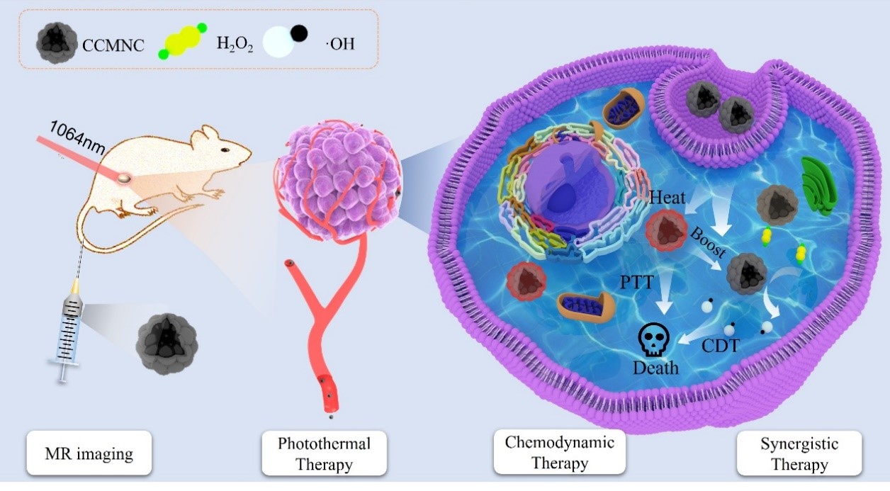
Schematic diagram of the CCMNCs for MR imaging, NIR-II-induced PTT-enhanced CDT.
They concluded that the CCMNCs could effectively absorb and convert NIR-II irradiation into cytotoxic heat, enhancing tumor CDT efficiency and producing synergistic therapeutic effects.
Read the original article on Chinese Academy of Sciences (CAS).