2025-07-13
Visited : 855
Low-cost, high-throughput method improves purity and reproducibility of EV-based proteomics.
A group of researchers at the VIB-UGent Center for Medical Biotechnology has developed a new platform to isolate and analyse extracellular vesicles (EVs), nanosized particles secreted by cells and playing a role in cellular communication and disease development. Called FAEVEr, the method increases the throughput of EV enrichment and is significantly more cost-efficient than existing methods. The study was published in the Journal of Extracellular Vesicles.
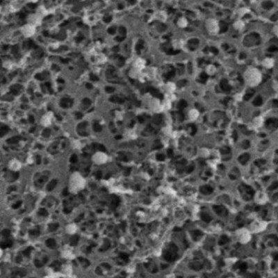
Extracellular Vesicles (EVs) are small particles that carry proteins, RNA, and other biomolecules from their cell of origin. They hold much promise for diagnostics and therapeutics, but isolating them from complex biofluids at high purity and throughput remains a major challenge. EVs are incredibly small - between 30 and 150 nanometers in size. To capture these tiny containers of messengers, scientists need to rely on sophisticated equipment such as ultracentrifuges. Unfortunately, these traditional methods of EV enrichment are time-consuming and resource-intensive with relatively low throughput.
Now, researchers in the lab of Prof. Kris Gevaert at the VIB-UGent Center for Medical Biotechnology have developed a novel EV-enrichment method. Building on their earlier work published in Molecular & Cellular Proteomics (January 2025), they demonstrated the effectiveness of Filter-Aided Extracellular Vesicle Enrichment, or FAEVEr in enriching EVs from individual samples in a miniaturized and 96-well high-throughput format.
Cost-efficient and fast
The new platform promises a much faster and more affordable alternative to existing EV-enrichment methods. Using 96-well filter plates, FAEVEr enables a much higher throughput volume by parallel processing of samples in under 2 hours.
Essential to the FAEVEr method is the use of Tween-20, a mild and inexpensive detergent added during EV washing steps. Its inclusion proved critical for improving the purity of the EV proteome by preventing fouling of the filter membranes used to trap EVs and by breaking weak interactions between EVs and contaminating abundant proteins. Importantly, this improvement in purity does not require complex chemistry or costly reagents, making the method accessible for both labs and clinical research settings using standard lab equipment.
Doing clinicians a FAEVEr
This innovation paves the way for more reproducible EV-based proteomics and strengthens the foundation for future clinical applications of EVs in diagnostics.
The method is already being applied in collaborations focused on prostate cancer, where enriched EV analysis may help identify disease-specific protein signatures from a simple urine sample.
Read the original article on VIB.