A team of researchers from US-based biosensor company SiPhox, alongside those from various US academic institutions, has used porous silicon (PSi) interferometer sensors to demonstrate the high contrast probe cleavage detection (HCPCD) mechanism.
The study, published on the preprint server bioRxiv, is the first of its kind, and acts as a “proof of principle” experiment, supporting the use of HCPCD as a sensitive and relatively low-cost method of providing point-of-care read-outs for a wide variety of diagnostic assays. This demonstration of the potential for HCPCD as a disposable point-of-care nucleic acid testing kit will likely be significant in the future management of the severe acute respiratory syndrome coronavirus 2 (SARS-CoV-2).
A Need for New Tests to Help Manage the Pandemic
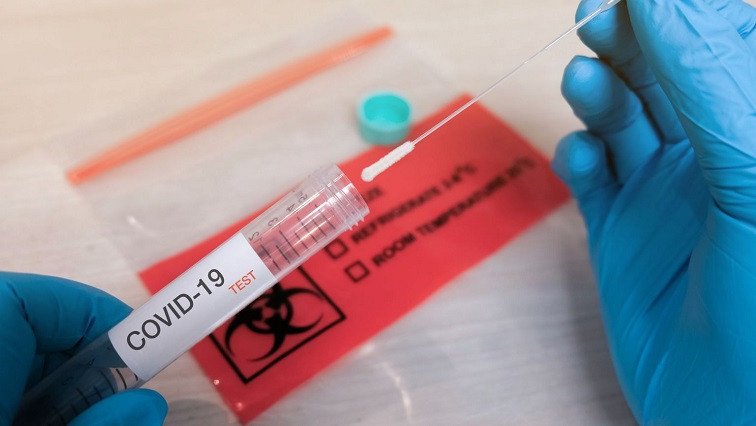
The scientific community is under pressure to make breakthroughs in nucleic acid testing to address the current need for COVID-19 tests and prepare the world for possible future pandemics.
Assays available for pathogen nucleic acid diagnostics have the drawback of needing to amplify the amount of ribonucleic acid (RNA) or deoxyribonucleic acid (DNA) of the target pathogen to be sensitive enough to detect the presence of the virus. These amplification processes are costly and time-consuming.
While photonic and plasmonic biosensing platforms offer a method of early-stage detection, they risk returning false negatives in the case where the level of viral RNA in the sample falls short of the sensor detection limit. These limits are defined by the number of molecules required to be captured for the sensor to detect them apart from the noise.
As new pathogens become more prevalent, developing new tests to address specific targets may take up to a year. As a result, scientists have explored how other methods can be developed to meet the need for accurate, sensitive, and rapid pathogen testing.
Previously, simulations have highlighted the potential of high contrast probe cleavage detection (HCPCD) as a sensitive diagnostic tool based on extracting probes from the photonic biosensor's surface.
Validating the Potential Diagnostic Applications of HCPCD
The researchers conducted DNase enzyme sensing experiments to confirm the ability of HCPCD to detect RNA and DNA, hence supporting its potential application in diagnostic testing. They made use of a porous silicon (PSi) interferometric sensor combined with high-contrast ds-DNA-quantum dots (Qdot) or low-index ds-DNA-fluorescein (FAM) probes.
The results demonstrated that while the attachment of Qdots achieved signal amplification and electrokinetic focusing, the approach was not suited to real-time diagnostics. On the other hand, HCPCD detection enhanced PSi sensors' sensitivity, highlighting the possibility of implementing HCPCD detection on silicon-based optical sensors. This would achieve the required sensitivity for a new pathogen nucleic acid diagnostic test and highlight the need for additional amplification methods.
Combining Psi Interferometer Sensors with HCPCD
In their paper, the project researchers outline the principle behind the technology. In the study, single layer PSi interferometers were used, which work on the thin film interference principle. The reflected light from the top and bottom interfaces of the sensor’s PSi layer interact, causing Fabry-Perot reflectance fringes. The positions of these fringes, as well as the periodicity of the interferometric pattern, are both a result of the PSi film’s adequate optical thickness.
Given that the wavelength of light is larger than the average pore diameter, scientists can use a valuable medium approximation to estimate the Psi film’s effective refractive index. Additionally, the chosen pore size is larger than the molecular target as well as the molecules of the surface functionalization process, allowing the large surface area to absorb the molecules. Once the molecules infiltrate the porous material's pores, this causes the effective refractive index of the PSi layer to increase and the fringes to redshift. By removing these molecules from the pores, the effective refractive index of PSi decreases, and the fringes blueshift.
Using HCPCD detection, the researchers first functionalized a PSi interferometer with high-index biotin-dsDNA-Qdot probes, resulting in a redshift of the reflectance fringes. Using an enzyme to cleave the DNA, the Qdots were removed, causing the fringes to blueshift. The reflectance shifts are directly proportional to the levels of the materials entering or exiting the pores, offering a method of optical readout that is both sensitive and quantitative.
Read the original article on AZoNano.