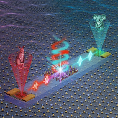
A new imaging technique, developed by the teams of Professors Jinyang Liang and Fiorenzo Vetrone at the Institut national de la recherche scientifique (INRS), can measure temperature in 2D, without contact, and in just a snap. The results of their research were published in the journal Nature Communications. This accurate real-time temperature detection could one day improve photothermal therapy and help in the early diagnosis of skin cancers.
This technology, known as single-shot photoluminescence lifetime imaging thermometry (SPLIT), is based on the luminescence of nanoparticles doped with rare earth ions. “These nanoparticles are considered as nanothermometers because their luminescent properties change with the temperature of the environment. They are also biocompatible,” says Professor Vetrone, a pioneer in this field of study.
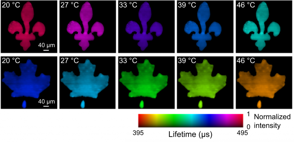
Lifetime images of green (top row) and red (bottom row) upconversion emission bands under different temperatures captured by SPLIT.
Instead of imaging the luminescence point by point, which is time consuming, SPLIT uses a novel ultrahigh-speed camera to track how quickly the luminescence decays of these nanoparticles in every spatial point.
The temperature can then be sensed by checking how fast the emitted light fades out. Since it is in real time, SPLIT can follow the phenomenon as it happens. For the first time, it enables the luminescence thermometry using the nanoparticle’s lifetime with a moving sample. “Compared to existing thermometry techniques, SPLIT is faster and has higher resolution. This allows a more accurate temperature sensing with both an advanced and economical solution,” adds Professor Liang, an expert in ultra-fast imaging.
Health applications
Professors Liang and Vetrone believe that SPLIT technology could, among other things, increase the ability to detect and treat skin cancers. At present, the capacity to detect melanomas, and more specifically micro-melanomas, is still limited. Existing diagnostic approaches are restricted by their invasiveness, resolution and accuracy, which leads to a large number of unnecessary biopsies.
Optical thermometry could thus be used to detect cancer cells, whose rapid metabolism leads to a higher temperature than that of normal tissue, making them more visible with SPLIT.
To detect melanoma, clinics can use a thermal camera, but the resolution is low. “SPLIT marks an important step in the technical development. With high resolution, the technology could be used to precisely locate the cancerous mole,” says Professor Liang.
Beyond detection, this technology could also be used to monitor the light dose during certain types of treatment. For example, photothermal therapy attacks cancer cells through the heat generated by exposure to near-infrared light. “We want to eradicate the cancer, but not the surrounding tissue, so if the temperature is too high, the treatment could be decreased or stopped for a while. If it’s too low, we can increase the light to get the right dose,” says Vetrone.
Read the original article on Institut national de la recherche scientifique (INRS).