Nanotechnology could help treat endometriosis, locating and removing painful, dangerous lesions in the ovaries, fallopian tubes and pelvis without invasive surgery, researchers from Oregon Health & Science University and Oregon State University found.
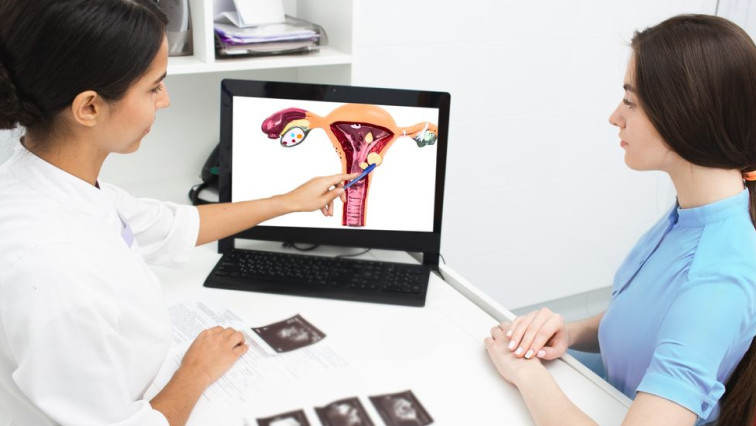
Endometriosis is a gynecological condition in which the endometrium tissue that normally lines the uterus forms lesions outside of the uterus. It causes infertility and severe pelvic pain in about 190 million women worldwide — roughly 10% of childbearing-age women. There is currently no cure.
Co-lead authors Ov Slayden, Ph.D., and Oleh Taratula, Ph.D., discussed their new paper detailing the approach on Oregon Public Broadcasting’s “Think Out Loud” program on April 29, the week following the paper’s publication in the journal Small.
Slayden, a professor in the OHSU Oregon National Primate Research Center, developed the animal model to test the iron oxide nanoparticles invented by Taratula, a professor in the OSU College of Pharmacy.
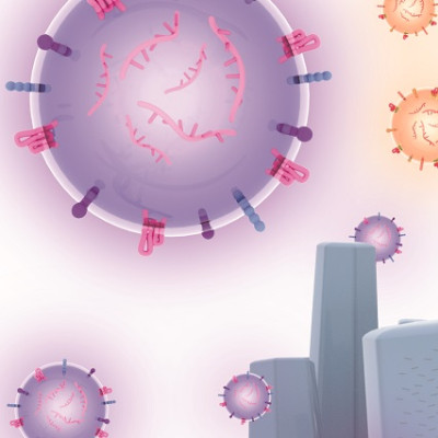
Iron oxide nanoparticles — the tiny particles about 1/1000th the size of a speck of dust — were injected intravenously into mice to locate lesions; they accumulated in the tissue lesions, enabling them to be identified by imaging.
Once the nanoparticles were in the lesions, they were exposed to an alternating magnetic field, causing the nanoparticles’ temperature to reach more than 120 degrees Fahrenheit — hot enough to remove the lesions with heat rather than surgery.
“Normally, treatment for endometriosis requires invasive surgical ablation of lesions. Lesions can return again and again, so that can mean multiple surgeries,” said Slayden, who has joint appointments in the OHSU School of Medicine departments of obstetrics and gynecology and physiology and pharmacology. “This treatment could provide a non-surgical option for treating the disease, which would be a dramatic quality-of-life improvement for the many millions of people who suffer from endometriosis.”
To test the nanoparticles, researchers transplanted endometriotic tissue from macaque primates into mice, to simulate endometriosis lesions.
The mice were injected with a low dose of nanoparticles that had been modified with a peptide specially designed to target a receptor that is abundant in endometriosis cells. By targeting that receptor, the nanoparticles were able to locate and accumulate in the lesions.
With the nanoparticles acting as a contrast agent spotlighting the lesions, OHSU radiologists Khashayar Farsad, M.D., Ph.D., associate professor of interventional radiology in the OHSU School of Medicine, and Cory Wyatt, Ph.D., research assistant professor of diagnostic radiology in the OHSU School of Medicine, were able to effectively image the lesions.
“Nanoparticle technology holds promise as a future molecular imaging tool in the diagnosis of many conditions, including endometriosis,” Farsad said. “Currently, small lesions are identified by direct visualization during surgery. Although very preliminary, this proof-of-concept study shows how nanotechnology could provide a novel non-invasive method to identify and potentially treat these lesions.”
To achieve a temperature hot enough to remove the lesions, OSU researchers developed nanoparticles that were hexagonal-shaped rather than spherical; the hexagonal shape has more than six times the heating efficiency once it’s exposed to an alternating magnetic field.
The research team’s next step is to apply their approach to a non-human primate model.
Read the original article on Oregon Health & Science University.