Medical imaging technology such as magnetic resonance imaging (MRI), computed tomography (CT) and X-ray could not provide real-time images during tumor resection surgery, and surgeons usually rely on visual, touch and experience to distinguish tumor margin, leading to a high probability of tumor residues.
Compared with traditional medical imaging, near-infrared (NIR) fluorescence imaging has the advantages of real-time, high sensitivity, high resolution, no radiation, etc. However, for tiny tumor residues, it requires relative high sensitivity to determine.
In a study published in Advanced Science, the research group led by Prof. ZHANG Yun from Fujian Institute of Research on the Structure of Matter of the Chinese Academy of Sciences developed a novel ultrasensitive nanoprobe (mZGS@Mn-AMD) with ultrahigh tumor-normal tissue (T/NT) signal ratio to distinguish residual tumor tissues from normal tissues.
Persistent luminescent nano core (mZGS) has high sensitivity because they do not need continuous excitation, and there is no background fluorescence interference generated by biological tissues during imaging.
The researchers quenched persistent luminescence (PersL) in normal tissue by the outer layer of MnO2, and recovered it due to the degradation of MnO2 in tumor microenvironment, improving the sensitivity of tumor imaging.
In orthotopic breast cancer model, the intraoperative tumor-to-normal tissue (T/NT) signal ratio of the nanoprobe was 58.8, about nine times that of downconversion nanoparticles. The T/NT ratio of residual tumor (< 2 mm) remained 12.4, considerably high to distinguish tumor tissue from normal tissue.
Besides, multiple-microtumor, 4T1 liver-implanted tumor and lung metastasis models were built to prove that this ultrasensitive nanoprobe is feasible to recognize tumor residues.
This study introduces an ultrasensitive and convenient imaging for recognizing residual tumor tissue, paving the way for complete surgical removal.
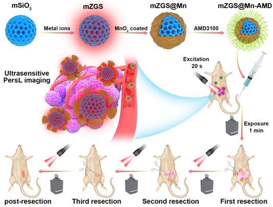
Schematic illustration of the Research.
Read the original article on Chinese Academy of Sciences (CAS).