This was revealed by a study, published on 3 January 2024 in the journal Nature Nanotechnology, conducted on glass nanofibers by a French-Chinese team including a CNRS chemist.
The reason for this is the inability of the macrophages naturally present in pulmonary alveolar tissue to eliminate foreign bodies that are too large. The study was initially conducted in vitro with electrochemical nanosensors, and revealed that when confronted with inert nanofibers over 15 microns in length, the cells are unable to distend enough to entirely encapsulate them within their “digestive” vesicle.
This results in leaked secretions that are very harmful for the alveolar walls, which this study detected, characterised, and quantified for the first time. An experiment on rats subsequently showed that regular unprotected inhalation of similar inert fibrous nanometerials, whatever they may be, causes repeated pulmonary lesions that can eventually lead to the development of fibroma.
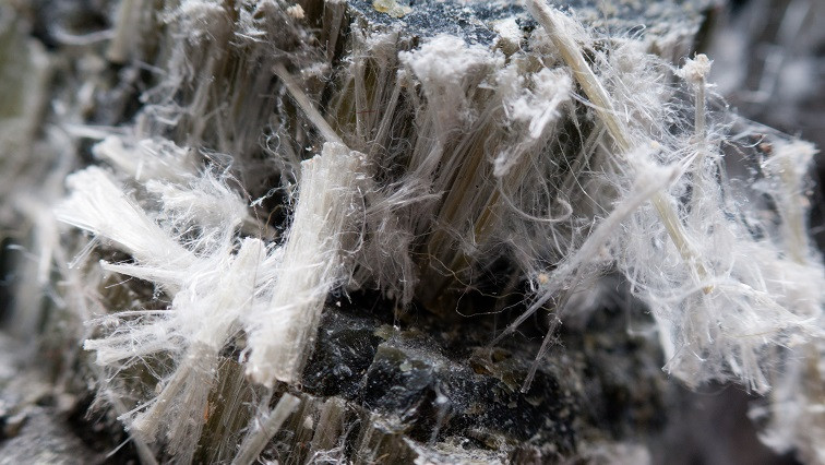
This discovery poses a challenge for the use of inert nanofibre felts in construction, which had heretofore been deemed to be less harmful than the asbestos it replaced, but that in reality could present the same health risks for those handling it.
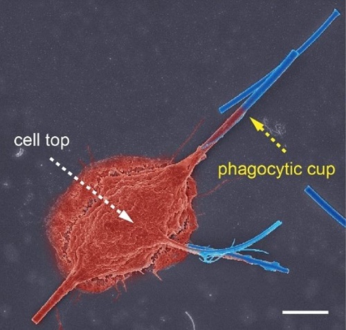
Pseudo-coloured scanning electron microscopy image: phagocytosis by a macrophage (red) of glass nanofibres (blue) after 12 h of frustrated phagocytosis; scale bar, 5 μm.
Read the original article on French National Centre for Scientific Research (CNRS).