Researchers often study biomolecules such as proteins or amino acids by chemically attaching a “fluorophore,” a sensitive molecule that absorbs and re-emits energy from light.
When activated by a laser and imaged through a high-powered microscope, these fluorophore tags or labels explode in a rainbow of color and information. They provide a wealth of insight that can, for example, help detect diseases or identify genetic conditions.
To detect more than one type of molecule at a time, or “multiplex” measurements, additional types of fluorophores that emit different colors of light are used. But it is surprisingly difficult to tell different colors apart at the single-molecule level. This is why most microscopes only look at three to four colors.
Researchers can break this color barrier using advanced techniques that involve days-long rounds of labeling and imaging or employ complicated setups with many lasers. Finding a simple and fast way to see many colors, however, has remained a major challenge.
Researchers at the UChicago Pritzker School of Molecular Engineering have a novel solution to this challenge, outlined in a paper published in Nature Nanotechnology. A new technique outlined by the Squires Lab uses three simple chemical building blocks to engineer dozens of “FRETfluor” tags, creating a more beautiful, nuanced spectrum of colors researchers can use to label biomolecules.
“Our approach is easier. It’s one shot of labeling, one shot of imaging,” said co-first author Jiachong Chu, a UChicago Pritzker Molecular Engineering PhD candidate. “That means you can do more with less. Currently, our novel technique is the best in the field.”
A new path toward multiplexing
Individual molecules are small and cell samples are comparatively huge, complicated and messy. The ultimate goal of this area of research – one that the PME team’s paper put closer than ever – is multiplexing.
“Multiplexing samples means to be able to, in the same measurement, measure more than one species of molecule so maybe you have 10 or 50, or hundreds of different proteins that you want to identify,” said Neubauer Family Assistant Professor of Molecular Engineering Allison Squires. “With this new technique, we can do dozens. I believe we can extend that to hundreds.”
To tackle this challenge, the Squires Lab team found an innovative new way to use a well-established technique: Förster Resonance Energy Transfer or FRET. FRET is a mechanism that describes how energy is transferred between light-sensitive molecules. It’s one way for researchers to measure the distance between different parts of a molecule, or to report when two molecules interact. FRET signals are exceptionally sensitive to the properties of the participating fluorophores, which the UChicago team used to tune their FRETfluor labels.
“This project utilizes FRET in a new way,” said co-first author Ayesha Ejaz, a PhD candidate in Chemistry. “FRET is commonly used for measuring distances and observing dynamics in biomolecules. We changed the spacing between a donor and acceptor dye to create different FRET efficiencies and other properties which we use to identify the different constructs.”
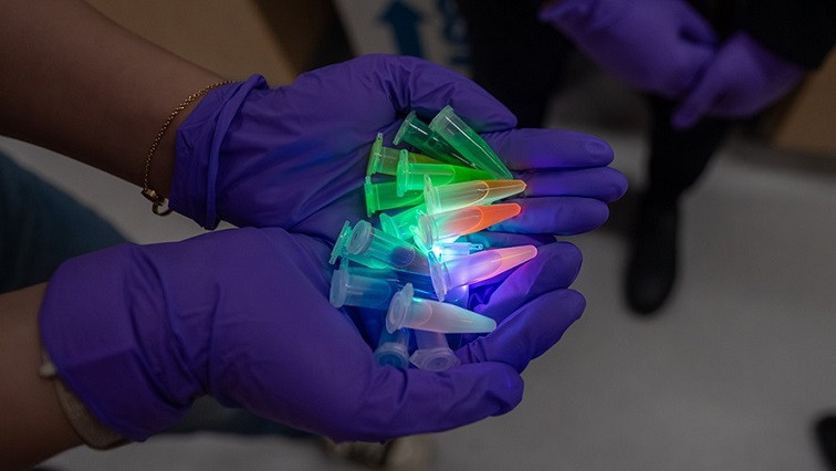
The 27 tags used in the PME team’s research were 27 “FRETfluors” they designed using a simple combination of DNA, a green cyanine dye (Cy3) and a red cyanine dye (Cy5). In addition to glowing in different colors, FRETfluors each exhibit other tunable properties such as the timing of how photons are emitted, or what the orientations of these photons are. Together, these properties can be used to identify a FRETfluor in just a fraction of a second, at ultra-low concentrations. Ejaz said one possible future direction for this research is to eventually replace ordinary fluorophore tags with these FRETfluors.
“Usually, when people want to look at multiple things – such as different parts of a cell – at once, they label each component with a different fluorescent tag that emits a certain color of light. But fluorescent tags are limited to four or five colors,” Ejaz said. “If FRETfluors can be used instead, then we can increase the number of ‘colors’ that are available for fluorescence microscopy. We are currently testing how well the FRETfluors work in different types of experiments and environments which will give us a better understanding of all the possibilities.”
“I’m excited to see the FRETfluors in action,” she said.
Sensitivity and simplicity
For Squires, much of the appeal of the new multiplexing technique comes from sensitivity combined with simplicity.
“Everybody wants to multiplex their favorite assay, and there are lots of existing strategies that will work in certain situations,” she said. “There are techniques that work well when you have tons of time, or when your sample is dead so that nothing moves. We're attacking the problem where you don't have tons of time. You want to know what disease somebody has while there’s still time to fight it, or you have only a teeny tiny bit of sample and you get one shot to identify each molecule as it flows through your channel. We can identify FRETfluors in a fraction of a second down to tens of femtomolar concentrations.”
Simplicity is key, both by using common chemicals to make the FRETfluors and by pioneering a technique that only needs one laser for readout.
“We only label to target once and only do the readout once,” Chu said. “Under that context, we can create 27 different tags that can be used at the same time.”
Squires described how existing techniques could be used together with FRETfluors for more multiplexing gains – “you could introduce fancy laser excitation schemes or incorporate other fluorophores that have slightly different properties” – that would improve the readouts from existing labels.
Applying these multipliers to their new, more powerful technique, Squires said, can open up worlds of new research and applications.
“These improvements to imaging and flow-based biomedical assays will enable the next generation of innovation,” Squires said.
Read the original article on University of Chicago.

