Key to the advance is a layer of gold added to devices that help visualise X-rays.
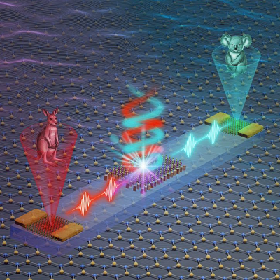
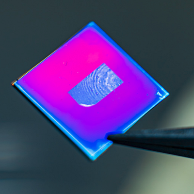
X-rays used in health and security scans are invisible, but they can be pictured using detectors that have “scintillating” materials which absorb the radiation and “light up” in a way similar to glow-in-the-dark paint. The visible light emitted by the scintillating materials is captured by sensors to create images based on the X-rays. The brighter the light, the sharper and more detailed the visuals.
The researchers, co-led by Nanyang Technological University, Singapore (NTU) and Poland’s Lukasiewicz Research Network-PORT Polish Centre for Technology Development, discovered that adding a gold layer to the scintillating materials made the visible light they gave off 120 per cent brighter. On average the light emitted had an intensity of around 88 photons per kiloelectronvolt, data from the study published in Advanced Materials showed.
As a result, the X-ray images produced were, in general, 38 per cent sharper and the ability to distinguish between different parts of the images was improved by 182 per cent.
With the gold layer, the time the scintillating materials took to stop emitting light after absorbing the X-rays was also shortened by 1.3 nanoseconds on average, or nearly 38 per cent, meaning they were ready for the next round of radiation more quickly. This suggests the potential for gold to speed up the processing of X-ray scans.
Rippling electrons
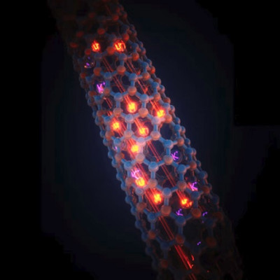
These boosts can be explained because gold is “plasmonic”, meaning the electrons in the metal react to radiation by moving in synchronised wave-like patterns, akin to ripples forming after a pebble is dropped into water.
These rippling electrons, also called plasmons, can interact with scintillating materials to accelerate the emission of visible light by the materials after they react with X-rays. This then causes the light given off to become more intense.
This contrasts with non-plasmonic materials, whose electrons do not interact with radiation in the same way. As a result, they do not move in a coordinated wave-like manner and do not speed up visible light emission by scintillating materials.
For the research, the experiments used gold just 70 nanometres thick, or about 1,000 times thinner than a strand of hair. Using a thin layer of gold helps to keep material costs down and keeps the size of future X-ray detectors compact.
The researchers added the plasmonic gold layer to a scintillating material called butylammonium lead bromide, from the “perovskite” family of compounds. Perovskites are known for their ability to convert sunlight into electricity in solar cells.
This “nanoplasmonic” study was conducted in collaboration between the CNRS-International-NTU-Thales Research Alliance, an NTU-based French-Singaporean joint research laboratory; Institut Lumière Matière CNRS based in Université Claude Bernard Lyon 1 in France; and Nano Center Indonesia.
Nanyang Assistant Professor Wong Liang Jie, study co-lead from NTU Singapore’s School of Electrical and Electronic Engineering, said: “Our results highlight the enormous potential of nanoplasmonics in optimising ultra-fast imaging systems where high spatial resolution and high contrast are needed, such as X-ray bioimaging and microscopy.”
Asst Prof Wong said that the improvements in X-ray detection demonstrated by the study stand to benefit airport security clearance too, as items in luggage might be more easily detected with crisper and higher-quality X-ray images, while bags could be screened more quickly.
Dr Muhammad Danang Birowosuto, study co-lead from the Lukasiewicz Research Network-PORT Polish Centre for Technology Development and a former NTU researcher, said: “Combining this improvement with other technologies will result in state-of-the-art functionalities in radiation imaging, such as to enhance X-ray analysis done in colour or improve the accuracy of ‘time-of-flight’ X-ray medical imaging.”
A spokesman for multinational corporation Thales said that “the idea of combining the physical phenomena of photonic structures – structures that change how light behaves – with scintillating materials for X-ray detectors represents an interesting concept to increase the efficiency of the current generation of detectors”.
“Thales continues to monitor scientific advances in this area with great interest and welcomes Asst Prof Wong’s breakthrough in this area,” the spokesman added.
Golden opportunity
The inspiration to use gold as a plasmonic material together with scintillating materials arose from a marriage of two research areas that had not been explored before for X-ray detectors.
Members of the research team previously found that after certain substances absorbed visible light, they also gave off visible light, which could get brighter if thin plasmonic gold at the nanometre scale was added.
At the time, other members of the team, who study how nano-sized structures enhance X-ray generation, were also working on X-ray detection.
Looking at the nanoplasmonic findings, an idea struck the team: Since X-ray detection in X-ray scanners also depends on substances absorbing radiation to emit visible light, could nanoscale plasmonic materials augment detectors in these scanners?
The scientists then set out to prove this experimentally with gold.
The researchers are next planning to add nano-sized notch-like patterns to the surface of the gold layer to boost the visible light given off by X-ray absorbing scintillating materials, as earlier research has shown that tiny notches can enhance visible light production.
Dr Dennis Schaart, head of the medical physics and technology section in the radiation science and technology department at the Netherlands’ Delft University of Technology, said that the findings “open a new avenue for the improvement of radiation imaging detectors based on scintillators”.
Scintillators convert X-ray or gamma-ray photons into measurable light signals for applications such as medical imaging in computed tomography (CT) scans, non-destructive testing like those for quality assurance in industrial production, and security clearance using airport baggage scanners.
Dr Schaart – who researches novel technologies for medical imaging and radiation oncology and was not involved in the study – said that the performance limits of commonly known scintillation mechanisms are close to being reached. But there remains a persistent demand for even better solutions.
“The findings presented in this latest research point the way towards a new class of scintillation detectors in which the intensity and speed of light emission are enhanced through the manipulation of quantum-mechanical phenomena,” he said.
“In principle, this offers highly exciting prospects for scintillator developers to engineer optimal materials for a wide variety of applications. If the results presented in the research can be reproduced and scaled towards industrially produced scintillators, this will likely contribute to, for example, more accurate, more affordable and more accessible medical diagnosis, as well as faster security scans.”
Read the original article on Nanyang Technological University (NTU).