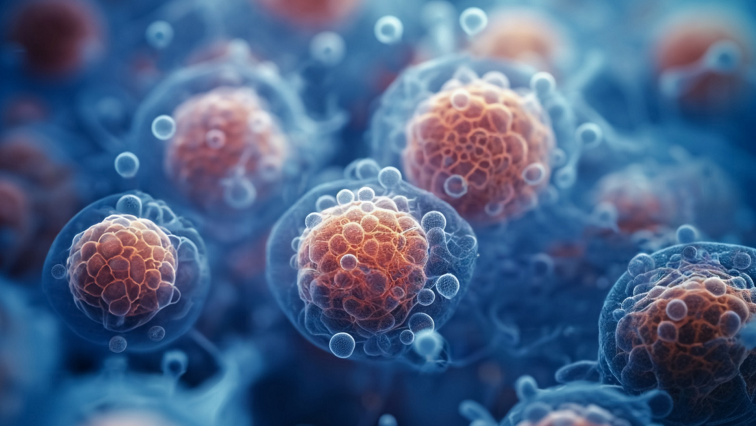
These particles, called extracellular vesicles, are considered attractive vehicle models for new drug therapies. But until now, researchers haven’t had the complete picture of how they work.
In a new study, a team led by medical researchers at The Ohio State University determined that these vesicles contain an ion channel – a protein that opens a corridor allowing electrical charges to pass through the protective outer membrane, a necessary step to keep contents and conditions stable inside.
Animal experiments also showed the ion channel influences the cargo, meaning the protein is important not just to the structure of extracellular vesicles (EVs), but also their function. Researchers compared the effects of RNA molecules delivered by EVs with and without the membrane protein to mice with ailing hearts. Only molecules carried by EVs with ion channels were able to repair the heart damage.
Harpreet Singh, professor of physiology and cell biology, and Mahmood Khan, professor of emergency medicine, both in Ohio State’s College of Medicine, co-led the study.
“We have not only discovered ion channels in these vesicles. We have recorded functional ion channels for the first time ever,” Singh said. “From forming a simple fundamental hypothesis that these vesicles should have ion channels all the way to showing that these vesicles will contain different cargo that can either protect or harm your cells – in this case, the heart – we have told the whole story.”
The paper was published Jan. 2 in Nature Communications.
Extracellular vesicles carry proteins and other molecules from donor to recipient cells to alter physiological and biological responses. In addition to facilitating cellular communication and maintaining cellular balance, the particles have been linked to immune responses, viral infectiousness, and cardiovascular disease, cancer and neurological disorders.
Based on his specialization in the study of ion channels, Singh predicted that EVs must have ion channels to safely transport molecules from cellular interiors to the extracellular environment and back into another type of cell. Otherwise, their membranes would be subject to bursting – caused by a rush of water triggered by osmotic stress or shock – as positive and negative electrical charges of ions in those varying environments ebb and flow.
“We know from our experience and from all this great work done in the last hundred years that ion channels are really, really important to maintain any structure which has a membrane,” Singh said.
Take the electrolyte potassium, for example. It is the most abundant positively charged ion inside cells, but its concentration is 30-fold lower in the extracellular environment.
“Suddenly an extracellular vesicle is coming from a huge potassium concentration to a low potassium concentration. What is going to happen if you can’t maintain ionic balance? You are going to feel the osmotic shock,” he said.
For this work, researchers isolated mouse EVs provided by Khan, also director of basic and translational research in the Department of Emergency Medicine, whose lab focuses on repairing damaged heart muscle with stem-cell therapy.
Because these particles are extremely small, the scientists created a technique they called near-field electrophysiology to record currents in the EV membranes. The method established the presence of a calcium-activated large-conductance potassium channel (BKCa).
They followed by isolating EVs from normal mice and knockout mice lacking the gene that encodes the BK potassium channel, and found the cargo in EVs from the knockout mice were very different in number and size – suggesting a functional role for the BKCa channel.
Several small RNA segments that regulate gene activation that were found among the cargo in the normal mouse vesicles were known to help protect the heart against oxidative stress, Khan said. EVs from the mice lacking the BK channel gene contained a different set of these segments, called microRNAs.
This finding led to the animal experiments in Khan’s lab, where EVs from normal mice and mice lacking the BK gene were injected into mice with diseased hearts.
“EVs from the wild-type animals protected the heart,” Singh said. “EVs that came out of the knockout mice could not protect the heart properly and, in fact, made things worse. Bad microRNAs were enriched in the vesicles that don’t have the channel.
“Is the cargo different because of different packaging, or is it because the vesicles without the channels are not surviving? That is an open question, and we are trying to address that.”
Another chief open question is identifying proteins, called transporters, that enable vesicles to maintain ionic balance as they transition from the extracellular environment back into a cell with a high potassium concentration.
Besides increasing fundamental knowledge about extracellular vesicles, Singh said, this work has potential to advance development of their use as therapeutics.
“People talk about loading these vesicles with charged molecules – whether it’s a drug, RNA proteins, or something else. If you’re loading them with charged molecules and you’re not managing ion homeostasis, you will have some sort of consequences,” he said. “That’s our big point, that if you are bioengineering EVs, you have to have the right combination of ion channels and transporters.”
Read the original article on Ohio State University.

