A great deal of time and money is required when developing medicines and vaccines. It is therefore crucial to be able to streamline the work by studying how, for example, individual proteins behave and interact with one another. The new microscopy method from Chalmers can enable the most promising candidates to be found at an earlier stage. The technique also has the potential for use in conducting research into the way cells communicate with one another by secreting molecules and other biological nanoparticles. These processes play an important role in our immune response, for example.
Revealing its silhouette
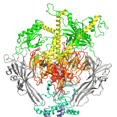
Biomolecules are both small and elusive, but vital since they are the building blocks of everything living. In order to get them to reveal their secrets using optical microscopy, researchers currently need to either mark them with a fluorescent label or attach them to a surface.
“With current methods you can never quite be sure that the labelling or the surface to which the molecule is attached does not affect the molecule’s properties. With the aid of our technology, which does not require anything like that, it shows its completely natural silhouette, or optical signature, which means that we can analyse the molecule just as it is,” says research leader Christoph Langhammer, professor at the Department of Physics at Chalmers. He has developed the new method together with researchers in both physics and biology at Chalmers and the University of Gothenburg.
The unique microscopy method is based on those molecules or particles that the researchers want to study being flushed through a chip containing tiny nano-sized tubes, known as nanochannels. A test fluid is added to the chip which is then illuminated with visible light. The interaction that then occurs between the light, the molecule and the small fluid-filled channels makes the molecule inside show up as a dark shadow and it can be seen on the screen connected to the microscope. By studying it, researchers can not only see but also determine the mass and size of the biomolecule, and obtain indirect information about its shape – something that was not previously possible with a single technique.
Acclaimed innovation
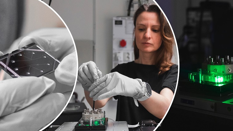
The new technique, nanofluidic scattering microscopy, was recently presented in the scientific journal Nature Methods. The Royal Swedish Academy of Engineering Sciences, which every year lists a number of research projects with the potential to change the world and provide real benefits, has also paid tribute to the progress made. The innovation has also taken a step out into society through the start-up company Envue Technologies, which was awarded the “Game Changer” prize in this year’s Venture Cup competition in Western Sweden.
“Our method makes the work more efficient, for example when you need to study the contents of a sample, but don’t know in advance what it contains and thus what needs to be marked,” says researcher Barbora Špačková, who during her time at Chalmers derived the theoretical basis for the new technique and then also conducted the first experimental study with the technology.
The researchers are now continuing to optimise the design of the nanochannels in order to find even smaller molecules and particles that are not yet visible today.
“The aim is to further hone our technique so that it can help to increase our basic understanding of how life works, and contribute to making the development of the next generation medicines more efficient” says Langhammer.
More about the scientific article and the research:
– The article Label-Free Nanofluidic Scattering Microscopy of Size and Mass of Single Diffusing Molecules and Nanoparticles was published in Nature Methods, and was written by Barbora Špačková, Henrik Klein Moberg, Joachim Fritzsche, Johan Tenghamn, Gustaf Sjösten, Hana Šípová-Jungová, David Albinsson, Quentin Lubart, Daniel van Leeuwen, Fredrik Westerlund, Daniel Midtvedt, Elin K. Esbjörner, Mikael Käll, Giovanni Volpe and Christoph Langhammer. The researchers are active at Chalmers and the University of Gothenburg. Barbora Špačková is currently starting up her own research group at the Czech Academy of Sciences in Prague.
– The research has been mainly funded by the Swedish Foundation for Strategic Research, as well as by the Knut and Alice Wallenberg Foundation. Part of the research was conducted at the Chalmers Nanofabrication Laboratory at the Department of Microtechnology and Nanoscience (MC2) and under the umbrella of the Chalmers Excellence Initiative Nano.
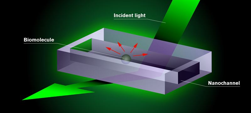
New microscopy method. The biomolecules the researchers want to study are placed in a chip consisting of tiny nano-sized tubes – nanochannels. Test fluid is added to the chip, which is mounted in an optical dark-field microscope and illuminated with visible light. The molecule appears as a dark shadow moving freely inside the channel on a screen connected to the microscope. The darkness of the shadow is proportional to the mass of the molecule. This is the result of an interference effect in the interaction of the light with the nanochannel and the molecule.
How the technique works
– The molecules or particles that the researchers want to study are placed in a chip containing tiny nano-sized tubes, nanochannels, that are filled with test fluid.
– The chip is secured in a specially adapted optical dark-field microscope and illuminated with visible light.
– On the screen that shows what can be seen in the microscope, the molecule appears as a dark shadow moving freely inside the nanochannel. This is due to the fact that the light interacts with both the channel and the biomolecule. The interference effect that then arises significantly enhances the molecule’s optical signature by weakening the light just at the point where the molecule is located.
– The smaller the nanochannel, the greater the amplification effect and the smaller the molecules that can be seen.
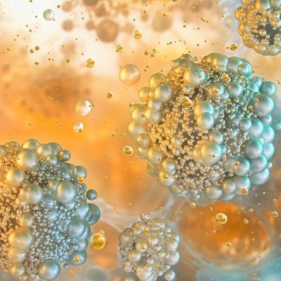
– With this technique it is currently possible to analyse biomolecules from a molecular weight of around 60 kilodaltons and upwards. It is also possible to study larger biological particles, such as extracellular vesicles and lipoproteins, as well as inorganic nanoparticles.
Read the original article on Chalmers University of Technology.