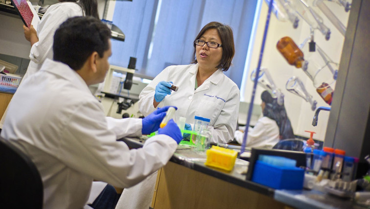
Kytai Nguyen, a UT Arlington bioengineering professor, is the principal investigator in the four-year, $2.1 million National Institutes of Health (NIH) grant. She’s collaborating with Jian Yang, a Penn State University bioengineering professor and former UTA faculty member, and Ralph Mason, a professor of radiology at UT Southwestern.
“What’s important in this project is that the technology carries fluorescent and ultrasound imaging capabilities, which will provide patients and doctors with more detailed information,” Nguyen said. “It also gives patients more targeted medicine, making it more efficient.”
PAD, more commonly known as either atherosclerosis or hardening of the arteries, is a condition commonly found in the elderly. It affects more than 200 million people worldwide and is associated with high rates of morbidity and mortality.
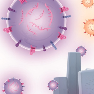
The research aims to develop novel biodegradable nanoparticles to deliver therapeutic agents that specifically protect cells under stress conditions, facilitate the formation of blood vessels under hypoxia and allow noninvasive multimodal imaging methods.
One impact of the research is to use these new nanoparticle platforms to deliver any therapeutics locally, treat the disease effectively and monitor the treatment noninvasively by imaging. The overall goal is to reduce complications and improve the quality of life for PAD patients, Nguyen said.
Michael Cho, chair of the UT Arlington Bioengineering Department, said Nguyen’s innovative research could greatly help those who live with PAD.
“This cutting-edge technology has a chance to change our protocols on how to deal with atherosclerosis,” Cho said. “When you are able to target localized lesions for treatment, that is so much better for the patients and much less invasive than current treatment.”
Read the original article on University of Texas at Arlington.