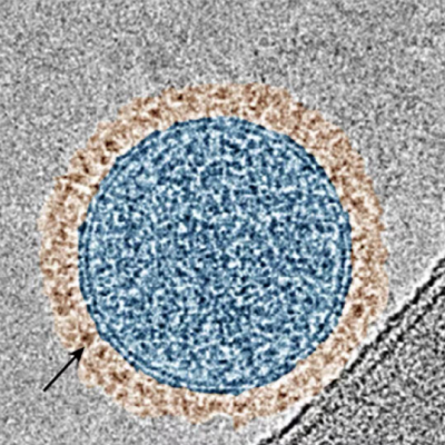
Adam J. Shuhendlernorth_eastexternal link, Associate Professor, at uOttawa’s Department of Chemistry and Biomolecular Sciences, and Scientist at the University of Ottawa Heart Institute, created these gold superclusters to work with the near-infrared light used in IV-OCT. These superclusters are made of tightly packed gold nanoparticles, which enhance the light scattering needed for clearer imaging.
"We've found a simple and quick way to produce these gold superclusters," says Shuhendler. "We can also adjust them to make them perfect for improving IV-OCT imaging."
The team coated the gold superclusters with a special polymer to stabilize them and allow targeting molecules to be attached.
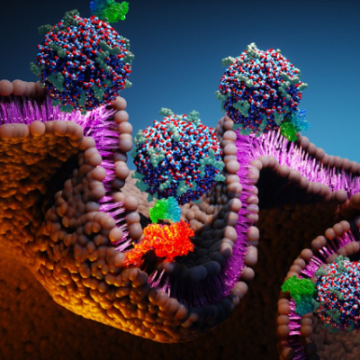
This study focused on P-selectin, a marker of blood vessel inflammation. The new contrast agent, named AuSC@(13FS)2, showed strong binding to P-selectin in lab tests and improved IV-OCT imaging in rats with inflamed blood vessels.
One major benefit of this new agent is that it can provide detailed molecular information without changing the existing IV-OCT procedures used in clinics. The researchers found that when AuSC@(13FS)2 bound to inflamed blood vessels, it created distinct reflections in the IV-OCT images, similar to those seen with stents.
"Our new contrast agent could lead to more personalized heart disease treatments," explains Shuhendler. "This technology might help doctors detect heart diseases earlier and assess the risk more accurately by providing detailed information about the blood vessels."
The study also showed a direct link between the amount of P-selectin and the number of reflections seen in the images, suggesting that this method could measure the severity of inflammation.
This research is a big step forward in heart disease imaging and diagnostics. By enabling detailed imaging with IV-OCT, it offers new opportunities for early detection and personalized treatment of heart conditions.
Read the original article on University of Ottawa.


