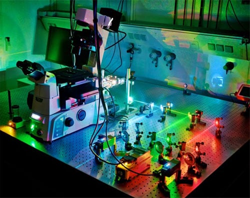
 With DNA-PAINT it is possible to visualize all the strands in DNA nanostructures individually. Courtesy of Maximilian Strauss, Max Planck Institute for Biochemistry.
DNA origami structures are essentially assembled by allowing one long single-stranded DNA molecule (the “scaffold” strand) to interact in a controlled, predefined manner with a set of shorter “staple” strands. The staple strands bind to specific stretches of the scaffold strand, progressively folding it into the desired form.
With DNA-PAINT it is possible to visualize all the strands in DNA nanostructures individually. Courtesy of Maximilian Strauss, Max Planck Institute for Biochemistry.
DNA origami structures are essentially assembled by allowing one long single-stranded DNA molecule (the “scaffold” strand) to interact in a controlled, predefined manner with a set of shorter “staple” strands. The staple strands bind to specific stretches of the scaffold strand, progressively folding it into the desired form.
The DNA-PAINT technique, developed by researchers at Ludwig Maximilian University (LMU), makes use of the specificity of DNA-DNA interactions. Short “imager” strands, linked to dye molecules that pair up with complementary sequences, are used to identify sites that are accessible for binding. Imager strands interact with their target sites, producing a blinking signal.
“By comparing the information in the individual fluorescence images, we are able to attain a higher resolution, so that we can inspect the whole structure in detail,” said Maximilian Strauss, researcher at the Max Planck Institute for Biochemistry at LMU.
“This phenomenon can be understood as follows," Strauss explained. "Let’s say we’re looking at a house with two illuminated windows. Seen from a certain distance, it appears as if the light is coming from one source. However, one can readily distinguish between the positions of the two windows if the lights are alternately switched on and off.”
In a similar way, the DNA-PAINT technique allows researchers to determine the positions of the bound staple strands precisely. The blinking signal emitted by imager strands reveals sites that are available for binding.
Use of DNA-PAINT revealed that strand incorporation strongly correlated with the position in the structure.
The DNA-PAINT method offers direct feedback for the rational refinement of the design and assembly process of DNA nanostructures and provides a quantitative explanation for efficiencies of DNA-based nanomachines.
According to researchers, the method can be applied to virtually any DNA-nanostructure geometry. Researchers believe that DNA-PAINT also could be used to characterize the labeling efficiency of antibodies or cellular proteins and nucleic acids, making it of interest for superresolution microscopy in general and quantitative structural biology in particular.
The research was published in Nature Communications (doi:10.1038/s41467-018-04031-z).
