
| Date | 12th, Apr 2018 |
|---|
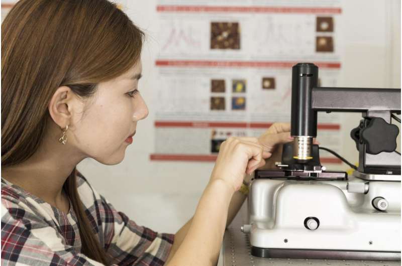
 Ph.D. student Sally Yunsun Kim works on the AFM-IR device in the Faculty of Pharmacy. Credit: Louise Cooper/University of Sydney
Researchers at the University of Sydney have established a method to identify individual nanoparticles released by human cells, opening the way for them to become diagnostic tools in the early-detection of cancers, dementia and kidney disease.
The particles, known as extracellular vesicles, or EVs, are routinely released by cells and play a central role in cell communication, sharing vital information such as DNA, RNA and proteins.
"This really is at the cutting edge of our knowledge of cellular development," said Associate Professor Wojciech Chrzanowski, co-author of a new paper on EVs published in the Royal Society of Chemistry's Nanoscale Horizons.
"EVs could not only be used to identify cellular pathologies but because they carry essential information about cell development, we could engineer them for purposes of tissue repair."
Associate Professor Chrzanowski from the University of Sydney Nano Institute and the Faculty of Pharmacy said the ability to identify individual EVs will provide biomarkers for diverse diseases such as cancers, cardiovascular, kidney and liver disease as well as dementia and multiple sclerosis.
He said it will also allow scientists to engineer EVs for use in tissue regeneration and help start a new chapter in stem-cell therapies and regenerative medicine.
"The human body naturally directs EVs from stem cells to damaged tissue to assist in its repair. By harnessing this knowledge, we could create a new generation of cell therapies," said Associate Professor Chrzanowski, who is the industry theme leader for Health and Medicine at Sydney Nano.
Understanding the particular nature of EVs is therefore essential for developing their application for diagnostics and therapeutics. For instance, early-stage cancerous cells release EVs that indicate the presence of malignant tissue in the body.
The study of extracellular vesicles is a relatively new field. It is only in the past decade that it has been known that cells communicate and transfer molecular and genetic information using EVs.
 Atomic force microscopy image of extracellular vesicles, scale bar at 200 nanometers. Credit: University of Sydney
The full potential to harness this knowledge for biomedical use has been hampered due to difficulties in establishing the heterogeneous nature of EV populations. Until now, they have only been analysed as large-scale populations with insufficient sensitivity.
Lead author of the paper, doctoral candidate Sally Yunsun Kim, said: "To unlock the true potential of EVs, what is needed is a new approach to unequivocally define nanoscale differences at a single EV level - and that is what we have done."
This is because it is the individual nature of the EVs as released by cells - affected by cellular morphology, genetics and environment - that give them their agency in human tissue repair.
Ms Kim, Associate Professor Chrzanowski and their team have developed a way to identify individual EV nanostructures, through examination of human placental stem cells provided by co-author Dr Bill Kalionis from the Royal Women's Hospital in Melbourne.
In the Nanoscale Horizons paper the team details a new method to identify the nanoscale composition of EVs using "resonance-enhanced atomic force microscope infrared spectroscopy" (AFM-IR).
This involves isolating singular EVs, thermally agitating them and then reading the particular signal or 'fingerprint' from this thermal activity using a 20-nanometre-wide detector.
Ms Kim, said: "We can do this using small amounts of human material, such as blood or urine samples. When cells create EVs they are spread throughout the body."
Associate Professor Chrzanowski said this ability to determine the particular nature of EVs will also allow scientists to continue fundamental research into how and why EVs are created by cells.
"This is a new and exciting field for biomedical research. And Australia is playing a leading role in this area," said Associate Professor Chrzanowski, who is a joint organiser of an international conference on extracellular vesicles that will take place in Sydney in November.
"The best people in the world will be here sharing their knowledge in a field with such promise for biomedical treatments," he said.
More information: Sally Yunsun Kim et al, None of us is the same as all of us: resolving heterogeneity of stem cell-derived extracellular vesicles using single-vesicle, nanoscale characterization with high-resolution resonance enhanced atomic force microscope infrared spectroscopy (AFM-IR), Nanoscale Horizons (2018). DOI: 10.1039/C8NH00048D
Citation: Scientists unlock path to use cell's own nanoparticles as disease biomarkers (2018, April 12) retrieved 22 August 2022 from https://phys.org/news/2018-04-scientists-path-cell-nanoparticles-disease.html
This document is subject to copyright. Apart from any fair dealing for the purpose of private study or research, no part may be reproduced without the written permission. The content is provided for information purposes only.
