Ground-breaking technology unlocks rapid and simple label-free 3D and 4D live cell imaging
A novel optical microscope utilizing diffraction tomography to generate 3D holographic images of unlabeled live cells is now available from Tomocube. Called HT-1, the new microscope delivers quantitative nanoscale, real-time, dynamic images of individual living cells quickly and simply without any sample preparation.
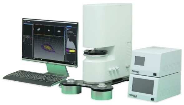
The new Tomocube HT-1 holotomography microscope, shown with its maglev-based antivibration TomoPlate and custom-made controllers for temperature and gas.
The holotomography images also deliver vital information on unique cell properties, including cell volume, shapes of sub-cellular organelles, cytoplasmic density, surface area, and deformability. The simplicity of operation and elimination of any preparative requirement will allow more researchers and clinicians to quantitatively study cell pathophysiology and the efficacy of drug treatments.
“Unlike expensive atomic force, electron and laser-scanning light microscopes that require extensive sample preparation and/or labeling, the HT-1’s 3D imaging technology relies on the same fundamental property of light as traditional phase contrast microscopes,” says Aubrey Lambert, Tomocube’s Chief Marketing Officer. “When light passes through any object, a phase shift is observable depending on its refractive index (RI). If this emergent light is viewed together with the original light, brightness changes can be seen dependent on the degree of phase shift. In the Tomocube microscope, a 3D image, or tomogram, is created from the RI measured at each 3-dimensional location during a 360-degree rotation. A similar concept using X-rays and nuclear magnetic moments is found in CT and MRI scanners.”
At the heart of the HT-1 is a complex digital micromirror device (DMD) optical light shaper, consisting of several hundred thousand micromirrors arranged in a rectangular array. Each individual mirror can be tilted plus or minus 12 degrees and the use of the DMD eliminates the need for moving parts in the lightpath. This patented technology allows the HT-1 to set its control beam rotation and rapidly switch its illumination angle to generate the 3D RI tomogram of the sample.
All the functions of the HT-1 are controlled by the TomoStudio software suite. A flexible user interface provides fast imaging capability and 2D/3D/4D visualization of the cellular images based on 3D RI distributions of the cells and tissues.
According the Lambert:
Cellular analysis plays a crucial role in a wide variety of research and diagnostic activities in the life sciences although the information available is limited by current microscopy techniques. The Tomocube HT-1 overcomes many of these limitations and open new vistas for researchers and clinicians to understand, diagnose and treat human diseases.”
