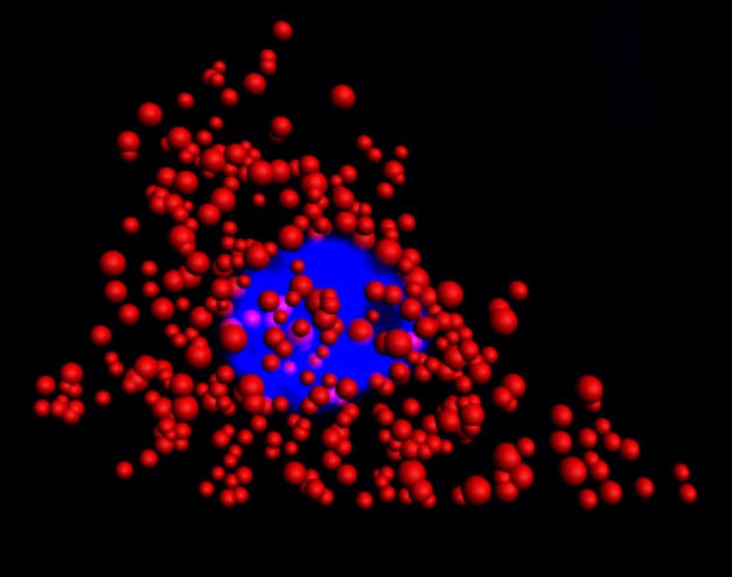
 Quantum dots illuminate the locations of individual mRNA as red dots in the cytoplasm of a single HeLa cell. The blue region is the nucleus. This work was a collaborative effort between Illinois Bioengineering and Mayo Clinic researchers. Courtesy of the University of Illinois at Urbana-Champaign Department of Bioengineering.
Signals from organic dye labels used in FISH rapidly deteriorate during photoexcitation, particularly when imaging in 3D and under high photon flux needed for superresolution. In addition, when dyes are used, the technique is limited to simultaneous analysis of about three RNA targets at a time.
Quantum dots illuminate the locations of individual mRNA as red dots in the cytoplasm of a single HeLa cell. The blue region is the nucleus. This work was a collaborative effort between Illinois Bioengineering and Mayo Clinic researchers. Courtesy of the University of Illinois at Urbana-Champaign Department of Bioengineering.
Signals from organic dye labels used in FISH rapidly deteriorate during photoexcitation, particularly when imaging in 3D and under high photon flux needed for superresolution. In addition, when dyes are used, the technique is limited to simultaneous analysis of about three RNA targets at a time.
Until now, size thresholds have limited the use of QDs for mRNA labeling in cells. To overcome this problem, the researchers identified the optimal size that QDs should be in order to effectively work with the FISH protocol. This discovery led them to develop QD-based FISH that matched the labeling accuracies currently obtained with organic dyes. The team created QDs made of a zinc, selenium, cadmium, and mercury alloy and coated with polymers.
“The core of the dot dictates the wavelength of emission, and the shell dictates how much light will be given off,” said professor Andrew Smith.
In experiments with HeLa cells and prostate cancer cells, the researchers found that dye-based FISH cell counts declined rapidly in minutes. The QD-based FISH method provided long-term luminescence to allow counting of RNA for more than 10 minutes, making it possible to acquire 3D cell imaging.
“This is important because images of cells and tissues are acquired slice-by-slice in sequence, so later slices that are labeled with dyes are depleted before they can be imaged,” Smith said.
The researchers believe that QD-FISH could particularly benefit high-resolution gene expression studies in 3D biological specimens for which quantification and multiplexing are major challenges.
“By replacing dyes with quantum dots, there are no stability issues whatsoever and we can count numerous RNAs with higher fidelity than before,” Smith said. “Moreover, we uncovered a fundamental limit to the size of a molecular label in cells, revealing new design rules for analysis in cells.”
This research was part of the Mayo Illinois Alliance in which engineers from Illinois work directly with clinicians and biologists from Mayo Clinic to solve outstanding medical challenges. The Mayo Clinic Biomarker Discovery Group is working to develop FISH-based diagnostics for tumor biopsies to improve the accuracy of cancer diagnosis, select personalized treatments, and improve prognoses.
The research was published in Nature Communications (https://doi.org/10.1038/s41467-018-06740-x).
