Ultrasensitive nanoscale optical probes have been created by scientists from UC Santa Cruz, to observe the bioelectric activity of neurons and other excitable cells.
 UC Santa Cruz engineers Ahsan Habib (left) and Ali Yanik have developed ultrasensitive nanoscale optical probes to monitor the bioelectric activity of neurons and other electrogenic cells. (Image credit: C. Lagattuta)
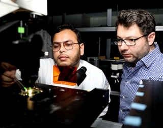
This innovative readout technology could allow researchers to analyze how neural circuits operate at a scale like never before, by observing large numbers of individual neurons at the same time. It could also pave the way for high-bandwidth brain-machine interfaces with drastically increased functionality and precision.
Traditionally, the electrical activity of neurons is monitored with the help of microelectrode arrays. However, these components are challenging to implement at a large scale and provide limited spatial resolution.
According to Ali Yanik, assistant professor of electrical and computer engineering at UCSC’s Baskin School of Engineering, the electronic wiring needed for readout is a key limitation of microelectrodes.
The extremely limited bandwidth of the electronic wiring is a bottleneck created by the very nature of electrons. We turn to photons because light offers billion-fold enhanced multiplexing and information carrying capabilities, the same reason why the telecommunication industry moved to fiber optics. By converting bioelectric signals to photons, we will be able to transmit large-bandwidth neural activity optically.
Ali Yanik, Assistant Professor of Electrical and Computer Engineering, Baskin School of Engineering, UC Santa Cruz
Collaborating with colleagues from the University of Notre Dame, Yanik’s lab has created extracellular nanoprobes that facilitate ultrasensitive optical monitoring of electrophysiological signals. Other techniques for performing optical monitoring necessitate genetic modifications to inject fluorescent molecules into cell membranes, which inhibits their use in humans.
Yanik’s technique is similar to extracellular microelectrode techniques, but only that the probes have nanoscale dimensions and the readout mechanism is optical. Furthermore, it produces a considerably brighter signal and higher signal-to-noise ratios compared to fluorescence-based probes.
“Harnessing the unparalleled multiplexing and information-carrying capability of light to dissect the neural circuitry and decrypt electrophysiological signals has been a goal of neuroscientists for nearly 50 years. We may have finally found a way to do that,” stated Yanik.
The new technology has been explained in a paper published in Science Advances on October 18th, 2019. The first author of the paper is Ahsan Habib, a PhD candidate in Yanik’s lab.
Broad Applications
The technology is still in its infancy, but according to Yanik, it could pave the way for a broad array of applications. Eventually, it could lead to robust brain-machine interfaces, allowing new brain-controlled prosthetic technologies to be developed for people with disabilities.
The optical nanoprobes developed by Yanik are nanoscale devices (with a diameter of
