May 27, 2020
(Nanowerk News) For the first time, scientists have introduced minuscule tracking devices directly into the interior of mammalian cells, giving an unprecedented peek into the processes that govern the beginning of development.
This work on one-cell embryos is set to shift our understanding of the mechanisms that underpin cellular behaviour in general, and may ultimately provide insights into what goes wrong in ageing and disease.
The research team included a trans-disciplinary partnership between embryologists in Bath,UK and the USA led by Professor Perry, and materials scientists and physicists led by Professor José Antonio Plaza at the Instituto de Microelectrónica de Barcelona (IMB-CNM) in Spain, involved injecting a silicon-based nanodevice together with sperm into the egg cell of a mouse. The result was a healthy, fertilised egg containing a tracking device (Nature Materials, "Tracking intracellular forces and mechanical property changes in mouse one-cell embryo development").
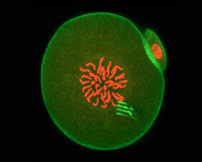 Fluorescence of an embryo containing a nanodevice. (Image: University of Bath)
The tiny devices are a little like spiders, complete with eight highly flexible ‘legs’. The legs measure the ‘pulling and pushing’ forces exerted in the cell interior to a very high level of precision, thereby revealing the cellular forces at play and showing how intracellular matter rearranged itself over time.
The nanodevices are incredibly thin – similar to some of the cell’s structural components, and measuring 22 nanometres, making them approximately 100,000 times thinner than a pound coin. This means they have the flexibility to register the movement of the cell's cytoplasm as the one-cell embryo embarks on its voyage towards becoming a two-cell embryo.
“This is the first glimpse of the physics of any cell on this scale from within,” said Professor Perry. “It’s the first time anyone has seen from the inside how cell material moves around and organises itself.”
Fluorescence of an embryo containing a nanodevice. (Image: University of Bath)
The tiny devices are a little like spiders, complete with eight highly flexible ‘legs’. The legs measure the ‘pulling and pushing’ forces exerted in the cell interior to a very high level of precision, thereby revealing the cellular forces at play and showing how intracellular matter rearranged itself over time.
The nanodevices are incredibly thin – similar to some of the cell’s structural components, and measuring 22 nanometres, making them approximately 100,000 times thinner than a pound coin. This means they have the flexibility to register the movement of the cell's cytoplasm as the one-cell embryo embarks on its voyage towards becoming a two-cell embryo.
“This is the first glimpse of the physics of any cell on this scale from within,” said Professor Perry. “It’s the first time anyone has seen from the inside how cell material moves around and organises itself.”
