The researchers assert that this has strong implications for not only electron microscopy, but also nanoscale technologies and various chip-based applications of analysis and sensing.
In their study, researchers noted that for their experiment to be successful, they needed to combine traveling-wave phase matching with a high density of photonic states. This was accomplished with the use of whispering-gallery mode microcavities.
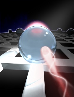
They used silica microspheres of 2 µm and 5 µm. Following excitation by a femtosecond pulse, the smaller sphere created stronger electron beams up to 700 eV, and the larger sphere allowed them to observe the coupling of the WGMs to the electrons.
Kfir said that with the light lasting for some time in the cavity, researchers were able to add additional laser energy to increase the effect.
“The efficient light-electron interaction can lead to a controllable optical control over regular electron microscopes, providing them with advancements such as higher sensitivity and time resolution,” he said.
In the future, the researchers, led by professor Claus Rogers, would use the coupling of electrons and WGMs to generate higher sensitivity and study the effects of excitation at the quantum level. “Using our unique ultrafast transmission electron microscope, such experiments can be combined with ultrafast temporal resolution and a spatial resolution of a nanometer, which could be amazing,” Kfir said.
This research was published in Nature (www.doi.org/10.1038/s41586-020-2320-y).
