Aug 26, 2020
(Nanowerk News) The study led by Imperial College London found that flexibility, as well as density, in the bone nanostructure is an important factor in assessing how likely someone is to suffer fractures.
The findings, published in Scientific Reports ("Nanoscale mechanisms in age-related hip-fractures"), suggest that doctors should look not only at bone density, but also bone flexibility, when deciding how to prevent bone breakages.
Imperial scientists test bone fragments under strain. On the right is its diffraction pattern.
Clinicians use DEXA scans, which look at how porous or dense bones are, to assess the likelihood of fractures. DEXA scans detect bone weakness in osteoporosis, a condition that causes weakened bones, and to inform treatments, like prescribing the medicine bisphosphonate, to help prevent fractures in these people.
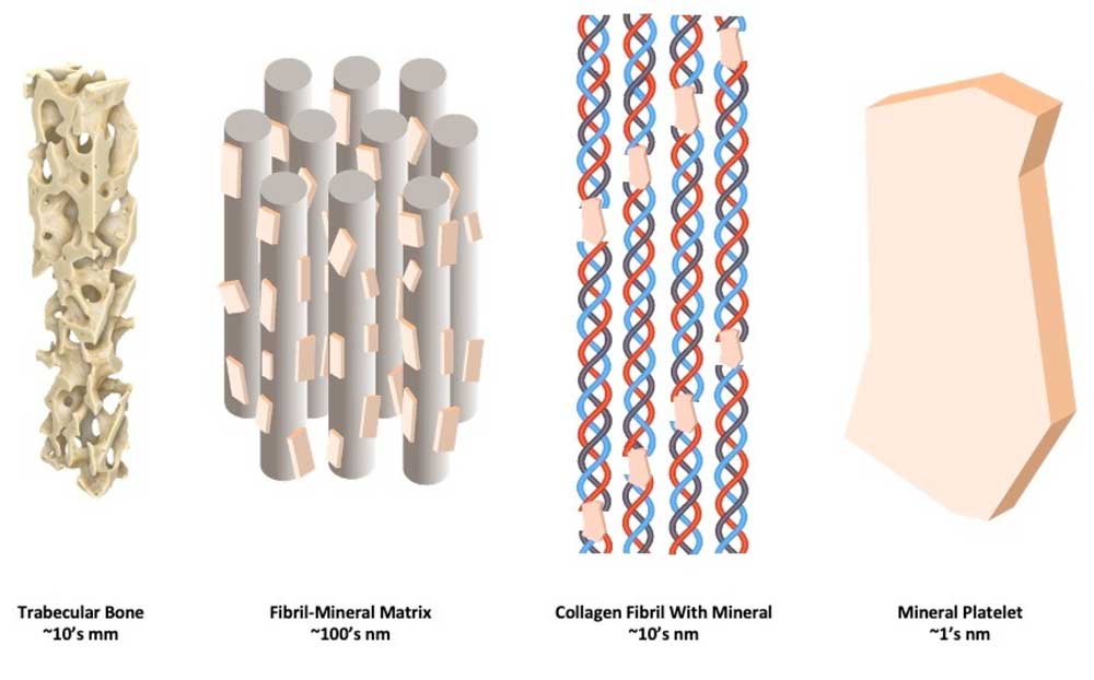
However, some people whose bones seem healthy on DEXA scans are more likely than others to suffer fractures. To find out why, the researchers looked to the building blocks of bone: stiff minerals surrounding flexible collagen fibrils, which are responsible for our bones' resistance to fracture during trips and falls.
They used high energy intense beams of X-rays generated by Diamond Light Source, the UK's national synchrotron, to examine bone flexibility at the nanoscale. They were able to assess how the collagen and minerals within bone flex and then break apart under load in the nanostructure of hip bone samples.
They compared the behaviour of the bone tissue samples under load between three groups of donors: those who had not suffered a hip fracture, or any other fracture; those without a bisphosphonate treatment history who had suffered a fractured hip; and those with a bisphosphonate treatment history who had suffered a fractured hip.
The team found that donors without fractures were more likely to have flexible collagen and mineral nanostructure than those with who had suffered fractures.
Irrespective of bisphosphonate treatment, the collagen and minerals were less flexible under load in patients with fractures, meaning the mineral broke away from the collagen at much lower forces.
The researchers say the bones may have fractured because the tissue was too inflexible and could not deform to absorb energy during a bump or fall - and that this highlights the importance of flexibility in the collagen and minerals of bone.
Therefore, flexibility at the nanoscale could be important in predicting future bone fractures and a target for new treatments - a finding that could inform future preventative treatment of bone fractures.
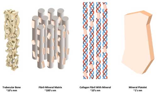 How stiff minerals and flexible collagen fibrils combine at the nanoscale to form bone. (Image: ICL) (click on image to enlarge)
Study co-author Dr Ulrich Hansen, of Imperial's Department of Mechanical Engineering, said: "We tend to think of our bones as solid, hard support structures, but flexibility appears to be extremely important in bone health. If bones are too hard, they are less able to absorb impact and more likely to break. Our study suggests that flexibility could be just as important as density in preventing fractures."
How stiff minerals and flexible collagen fibrils combine at the nanoscale to form bone. (Image: ICL) (click on image to enlarge)
Study co-author Dr Ulrich Hansen, of Imperial's Department of Mechanical Engineering, said: "We tend to think of our bones as solid, hard support structures, but flexibility appears to be extremely important in bone health. If bones are too hard, they are less able to absorb impact and more likely to break. Our study suggests that flexibility could be just as important as density in preventing fractures."
