| Date | 1st, May 2021 |
|---|
Home > Press > World's first fiber-optic ultrasonic imaging probe for future nanoscale disease diagnostics
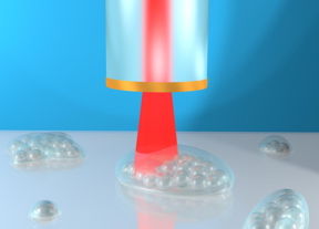 Concept art showing the 3D mapping of microscopic objects by the phonon probe system. The optical fibre contains a metal layer on its tip and projects red laser light into the specimen
CREDIT
Dr Salvatore La Cavera
Concept art showing the 3D mapping of microscopic objects by the phonon probe system. The optical fibre contains a metal layer on its tip and projects red laser light into the specimen
CREDIT
Dr Salvatore La Cavera
Abstract: Scientists at the University of Nottingham have developed an ultrasonic imaging system, which can be deployed on the tip of a hair-thin optical fibre, and will be insertable into the human body to visualise cell abnormalities in 3D.
Nottingham, UK | Posted on April 30th, 2021
The new technology produces microscopic and nanoscopic resolution images that will one day help clinicians to examine cells inhabiting hard-to-reach parts of the body, such as the gastrointestinal tract, and offer more effective diagnoses for diseases ranging from gastric cancer to bacterial meningitis.
The high level of performance the technology delivers is currently only possible in state-of-the-art research labs with large, scientific instruments - whereas this compact system has the potential to bring it into clinical settings to improve patient care.
The Engineering and Physical Sciences Research Council (EPSRC)-funded innovation also reduces the need for conventional fluorescent labels - chemicals used to examine cell biology under a microscope - which can be harmful to human cells in large doses.
The findings are being reported in a new paper, entitled 'Phonon imaging in 3D with a fibre probe' published in the Nature journal, Light: Science & Applications.
Paper author, Salvatore La Cavera, an EPSRC Doctoral Prize Fellow from the University of Nottingham Optics and Photonics Research Group, said of the ultrasonic imaging system: "We believe its ability to measure the stiffness of a specimen, its bio-compatibility, and its endoscopic-potential, all while accessing the nanoscale, are what set it apart. These features set the technology up for future measurements inside the body; towards the ultimate goal of minimally invasive point-of-care diagnostics."
Currently at prototype stage, the non-invasive imaging tool, described by the researchers as a "phonon probe", is capable of being inserted into a standard optical endoscope, which is a thin tube with a powerful light and camera at the end that is navigated into the body to find, analyse, and operate on cancerous lesions, among many other diseases. Combining optical and phonon technologies could be advantageous; speeding up the clinical workflow process and reducing the number of invasive test procedures for patients.
3D mapping capabilities
Just as a physician might conduct a physical examination to feel for abnormal 'stiffness' in tissue under the skin that could indicate tumours, the phonon probe will take this '3D mapping' concept to a cellular level.
By scanning the ultrasonic probe in space, it can reproduce a three-dimensional map of stiffness and spatial features of microscopic structures at, and below, the surface of a specimen (e.g. tissue); it does this with the power to image small objects like a large-scale microscope, and the contrast to differentiate objects like an ultrasonic probe.
"Techniques capable of measuring if a tumour cell is stiff have been realised with laboratory microscopes, but these powerful tools are cumbersome, immobile, and unadaptable to patient-facing clinical settings. Nanoscale ultrasonic technology in an endoscopic capacity is poised to make that leap," adds Salvatore La Cavera.
How it works
The new ultrasonic imaging system uses two lasers that emit short pulses of energy to stimulate and detect vibrations in a specimen. One of the laser pulses is absorbed by a layer of metal - a nano-transducer (which works by converting energy from one form to another) - fabricated on the tip of the fibre; a process which results in high-frequency phonons (sound particles) getting pumped into the specimen. Then a second laser pulse collides with the sound waves, a process known as Brillouin scattering. By detecting these "collided" laser pulses, the shape of the travelling sound wave can be recreated and displayed visually.
The detected sound wave encodes information about the stiffness of a material, and even its geometry. The Nottingham team was the first to demonstrate this dual-capability using pulsed lasers and optical fibres.
The power of an imaging device is typically measured by the smallest object that can be seen by the system, i.e. the resolution. In two dimensions the phonon probe can "resolve" objects on the order of 1 micrometre, similar to a microscope; but in the third dimension (height) it provides measurements on the scale of nanometres, which is unprecedented for a fibre-optic imaging system.
Future applications
In the paper, the researchers demonstrate that the technology is compatible with both a single optical fibre and the 10-20,000 fibres of an imaging bundle (1mm in diameter), as used in conventional endoscopes.
Consequently, superior spatial resolution and wide fields of view could routinely be achieved by collecting stiffness and spatial information from multiple different points on a sample, without needing to move the device - bringing a new class of phonon endoscopes within reach.
Beyond clinical healthcare, fields such as precision manufacturing and metrology could use this high-resolution tool for surface inspections and material characterisation; a complementary or replacement measurement for existing scientific instruments. Burgeoning technologies such as 3D bio-printing and tissue engineering could also use the phonon probe as an inline inspection tool by integrating it directly to the outer diameter of the print-needle.
Next, the team will be developing a series of biological cell and tissue imaging applications in collaboration with the Nottingham Digestive Diseases Centre and the Institute of Biophysics, Imaging and Optical Science at the University of Nottingham; with the aim to create a viable clinical tool in the coming years.
###
####
About University of NottinghamThe University of Nottingham is a research-intensive university with a proud heritage, consistently ranked among the world's top 100. Studying at the University of Nottingham is a life-changing experience and we pride ourselves on unlocking the potential of our students. We have a pioneering spirit, expressed in the vision of our founder Sir Jesse Boot, which has seen us lead the way in establishing campuses in China and Malaysia - part of a globally connected network of education, research and industrial engagement. The University's state-of-the-art facilities and inclusive and disability sport provision is reflected in its status as The Times and Sunday Times Good University Guide 2021 Sports University of the Year. We are ranked eighth for research power in the UK according to REF 2014. We have six beacons of research excellence helping to transform lives and change the world; we are also a major employer and industry partner - locally and globally. Alongside Nottingham Trent University, we lead the Universities for Nottingham initiative, a pioneering collaboration which brings together the combined strength and civic missions of Nottingham's two world-class universities and is working with local communities and partners to aid recovery and renewal following the COVID-19 pandemic.
For more information, please click here
Contacts:Emma Lowry
44-011-584-67156
More information is available from Salvatore La Cavera III on
@UoNPressOffice
Copyright © University of Nottingham
If you have a comment, please Contact us.
Issuers of news releases, not 7th Wave, Inc. or Nanotechnology Now, are solely responsible for the accuracy of the content.
Bookmark:


News and information
![]() Bumps could smooth quantum investigations: Rice University models show unique properties of 2D materials stressed by contoured substrates June 10th, 2022
Bumps could smooth quantum investigations: Rice University models show unique properties of 2D materials stressed by contoured substrates June 10th, 2022
![]() Emerging vaccine nanotechnology June 3rd, 2022
Emerging vaccine nanotechnology June 3rd, 2022
![]() �Fruitcake� structure observed in organic polymers June 3rd, 2022
�Fruitcake� structure observed in organic polymers June 3rd, 2022
![]() Progressive Medicinal and Herbal Nanoscience for Targeted Drug Delivery Systems June 3rd, 2022
Progressive Medicinal and Herbal Nanoscience for Targeted Drug Delivery Systems June 3rd, 2022
Imaging
![]() Snapshot measurement of single nanostructure�s circular dichroism March 25th, 2022
Snapshot measurement of single nanostructure�s circular dichroism March 25th, 2022
![]() Better understanding superconductors with Higgs spectroscopy Prof. Stefan Kaiser from TU Dresden awarded ERC Consolidator Grant March 18th, 2022
Better understanding superconductors with Higgs spectroscopy Prof. Stefan Kaiser from TU Dresden awarded ERC Consolidator Grant March 18th, 2022
![]() Visualizing the invisible: New fluorescent DNA label reveals nanoscopic cancer features March 4th, 2022
Visualizing the invisible: New fluorescent DNA label reveals nanoscopic cancer features March 4th, 2022
Possible Futures
![]() Bumps could smooth quantum investigations: Rice University models show unique properties of 2D materials stressed by contoured substrates June 10th, 2022
Bumps could smooth quantum investigations: Rice University models show unique properties of 2D materials stressed by contoured substrates June 10th, 2022
![]() Progressive Medicinal and Herbal Nanoscience for Targeted Drug Delivery Systems June 3rd, 2022
Progressive Medicinal and Herbal Nanoscience for Targeted Drug Delivery Systems June 3rd, 2022
![]() Artificial Intelligence Centered Cancer Nanomedicine: Diagnostics, Therapeutics and Bioethics June 3rd, 2022
Artificial Intelligence Centered Cancer Nanomedicine: Diagnostics, Therapeutics and Bioethics June 3rd, 2022
Nanomedicine
![]() Emerging vaccine nanotechnology June 3rd, 2022
Emerging vaccine nanotechnology June 3rd, 2022
![]() Progressive Medicinal and Herbal Nanoscience for Targeted Drug Delivery Systems June 3rd, 2022
Progressive Medicinal and Herbal Nanoscience for Targeted Drug Delivery Systems June 3rd, 2022
![]() Artificial Intelligence Centered Cancer Nanomedicine: Diagnostics, Therapeutics and Bioethics June 3rd, 2022
Artificial Intelligence Centered Cancer Nanomedicine: Diagnostics, Therapeutics and Bioethics June 3rd, 2022
![]() Nanostructured fibers can impersonate human muscles June 3rd, 2022
Nanostructured fibers can impersonate human muscles June 3rd, 2022
Discoveries
![]() Bumps could smooth quantum investigations: Rice University models show unique properties of 2D materials stressed by contoured substrates June 10th, 2022
Bumps could smooth quantum investigations: Rice University models show unique properties of 2D materials stressed by contoured substrates June 10th, 2022
![]() Emerging vaccine nanotechnology June 3rd, 2022
Emerging vaccine nanotechnology June 3rd, 2022
![]() �Fruitcake� structure observed in organic polymers June 3rd, 2022
�Fruitcake� structure observed in organic polymers June 3rd, 2022
Announcements
![]() Bumps could smooth quantum investigations: Rice University models show unique properties of 2D materials stressed by contoured substrates June 10th, 2022
Bumps could smooth quantum investigations: Rice University models show unique properties of 2D materials stressed by contoured substrates June 10th, 2022
![]() Progressive Medicinal and Herbal Nanoscience for Targeted Drug Delivery Systems June 3rd, 2022
Progressive Medicinal and Herbal Nanoscience for Targeted Drug Delivery Systems June 3rd, 2022
![]() Artificial Intelligence Centered Cancer Nanomedicine: Diagnostics, Therapeutics and Bioethics June 3rd, 2022
Artificial Intelligence Centered Cancer Nanomedicine: Diagnostics, Therapeutics and Bioethics June 3rd, 2022
Interviews/Book Reviews/Essays/Reports/Podcasts/Journals/White papers/Posters
![]() �Fruitcake� structure observed in organic polymers June 3rd, 2022
�Fruitcake� structure observed in organic polymers June 3rd, 2022
![]() Progressive Medicinal and Herbal Nanoscience for Targeted Drug Delivery Systems June 3rd, 2022
Progressive Medicinal and Herbal Nanoscience for Targeted Drug Delivery Systems June 3rd, 2022
![]() Artificial Intelligence Centered Cancer Nanomedicine: Diagnostics, Therapeutics and Bioethics June 3rd, 2022
Artificial Intelligence Centered Cancer Nanomedicine: Diagnostics, Therapeutics and Bioethics June 3rd, 2022
Tools
![]() Snapshot measurement of single nanostructure�s circular dichroism March 25th, 2022
Snapshot measurement of single nanostructure�s circular dichroism March 25th, 2022
![]() Eyebrow-raising: Researchers reveal why nanowires stick to each other February 11th, 2022
Eyebrow-raising: Researchers reveal why nanowires stick to each other February 11th, 2022
![]() JEOL Introduces New Scanning Electron Microscope with �Simple SEM� Automation and Live Elemental and 3D Analysis January 14th, 2022
JEOL Introduces New Scanning Electron Microscope with �Simple SEM� Automation and Live Elemental and 3D Analysis January 14th, 2022
![]() Super-resolved imaging of a single cold atom on a nanosecond timescale January 7th, 2022
Super-resolved imaging of a single cold atom on a nanosecond timescale January 7th, 2022
