A recent review published in the journal Nanoscale Research Letters explores the biomedical applications of fluorescent nanoparticles, including semiconductor quantum dots, silica nanoparticles, carbon nanotubes, and nanoparticles.
Study: Advances and Challenges of Fluorescent Nanomaterials for Synthesis and Biomedical Applications. Image Credit: Golden Wind/Shutterstock.com
Importance of Fluorescent Nano Materials
Fluorescent nanomaterials (FNMs) have superior optical features due to their nanoscale size, which is crucial for bioimaging applications and fluorescence-dependent detection techniques.
The physical and mechanical characteristics of fluorescent nanoparticles are determined by their geometry, structure, and composition, which significantly impact their effectiveness. Considerable research has been expended in previous years to increase the bioavailability of fluorescent nanoparticles by refining the production procedure.
Limitations in the Past
The limited specificity and photoluminescence effectiveness of fluorescent nanoparticles limit their applicability in imaging and detection to some extent.
Their uses in biological imaging and bio labeling were jeopardized due to their poor fluorescence and inherent cytotoxicity. Although, scientists are continuously working to synthesize new FNMs with improved features.
Various Types of Fluorescent Nanoparticles
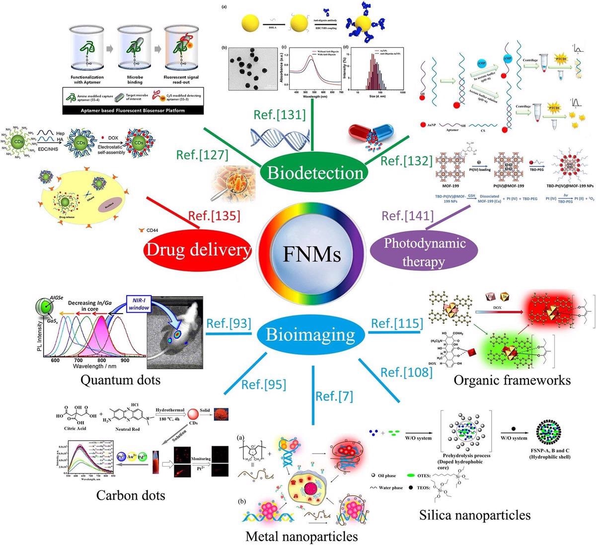
Keeping in mind their broad absorbance and homogeneous photoluminescence spectrum, fluorescence quantum yield, resilience to photo corrosion, higher molecular extinction factors, and relatively large Stokes adjustments, quantum dots (QDs) have been a popular study topic in recent decades.
In terms of the QD creation mechanism, when electric charges (electrons and holes) are limited to specific locations by potential barriers, a shift in spectra occurs, leading to their formation.
Carbon dots (CDs) are new nanoparticles in the carbon-based family with diameters smaller than 10 nm. Fluorescent carbon nanoparticles are gaining popularity for cell imaging and other medical applications due to their low toxicity, resilience to photo corrosion, and better bioactivity. Carbon nanoparticles have more than 20 nm diameters compared to the average sizes of carbon dots of 1–6 nm.
The overview diagram of biomedical applications of fluorescent nanomaterials. Image Credit: Xiao, D. et al.
Because of their electrical and optical characteristics, one-dimensional (1D) carbon nanotubes have sparked much interest in the healthcare field. According to the numbers of tubular graphene layers, carbon nanotubes are classified as single-walled carbon nanotubes (SWCNTs) or multi-walled carbon nanotubes (MWCNTs).
Graphene and its variants have been extensively researched as two-dimensional carbon nanomaterials for various biological purposes such as imaging and medication delivery. They have a higher surface area and distinct surface morphology that allow for electrostatic interaction with dye molecules.
When compared to QDs, noble metal atoms exhibit reduced toxicity. Gold, silver, and copper nanoparticles are gaining popularity and are being used in various applications. Silica nanoparticles are widely used in the biomedical industry due to their opacity, physical resilience, resilience, and stabilization of the implanted fluorophores.
Due to their inherent benefits in decreasing autofluorescence and light-dispersion interference from tissues, phosphors are frequently utilized in biomedical science. Phosphors, in general, are made up of compounds and doped ions.
COFs (covalent organic frameworks) are novel permeable crystal nanoparticles with high durability, adhesion, and low cytotoxicity. The creation of fluorescent tiny organic compounds combined with the fluorescence estimation process can be utilized to build more effective nanomachines.
Applications of FNPs
These nanomaterials are an attractive choice for biological applications. Fluorescent nanomaterials may dramatically increase fluorescence signals while being biocompatible, resulting in a growing interest in their use for fast monitoring of biomolecules.
Aside from disease detection, fluorescent nanoparticles have piqued the interest of investigators in DNA detection. Photodynamic therapy (PDT) has also been cited as a revolutionary tumor therapeutic strategy that makes use of the interplay between light and photocatalyst.
Challenges
With the aggregation of microscopic particles during the fabrication process, the dimension and shape dispersion of FNMs can rarely be distributed evenly, which might harm the optical characteristics of FNMs in biological applications.
As a result, FNM applicability is currently limited to the research size. Their fluorescence will be diminished or perhaps eliminated in the reaction mixture, or solid form, known as Aggregation Caused Quenching (ACQ). This dilemma has perplexed researchers for almost 150 years, preventing the widespread use of fluorescent dyes.
Autofluorescence decreases the signal-to-background proportion and frequently disrupts digital effects. Although using FNMs in mice produced satisfactory results, achieving equivalent results in other animal models remains problematic.
Restrictive aspects of traditional materials and procedures in biological applications can be overcome thanks to the unique features of fluorescent nanoparticles.
For biological applications, QDs with electro-optical components are appealing. Efficient biological imaging, biological sensing, and phototherapy will become more frequently used in illness management and prognosis as fluorescent nanomaterials evolve.
Continue reading: The Role of Titanium Nanostructures in Biomedical Applications.
Reference
Xiao, D. et al., (2021) Advances and Challenges of Fluorescent Nanomaterials for Synthesis and Biomedical Applications. Nano Research Letters, 16(17). Available at: https://nanoscalereslett.springeropen.com/articles/10.1186/s11671-021-03613-z
Disclaimer: The views expressed here are those of the author expressed in their private capacity and do not necessarily represent the views of AZoM.com Limited T/A AZoNetwork the owner and operator of this website. This disclaimer forms part of the Terms and conditions of use of this website.


