In a pre-proof paper available in the journal Biochemical and Biophysical Research Communications researchers demonstrated the key role played by the geometry of graphene oxide (GO) nanomaterials, such as quantum dots and nanosheets, in influencing the biophysical impacts on hepatic stellate cells (HSCs).
Study: The geometry-dependent regulation of hepatic stellate cells by graphene oxide nanomaterials. Image Credit: Jose Luis Calvo/Shutterstock.com
Graphene Oxide in Biomedical Applications
Nanomaterials are used extensively in biomedical applications such as photothermal therapy, bioimaging, and drug delivery. Specifically, graphene oxide (GO) nanomaterials are one of the most preferred drug delivery methods for the treatment of liver diseases owing to their biocompatibility and tunable physical and chemical properties. GO quantum dots and nanosheets are the common GO geometric forms used in biomedical applications.
However, a significant share of administered nanoparticles is often accumulated in livers for a considerable duration. The time needed for their clearance ranges between 1 and 21 days, based on the different physiochemical properties of nanomaterials. This long retention time and slow clearance can lead to chronic toxicity to livers and cause liver diseases in the long term, such as liver fibrosis.
Liver fibrosis is primarily triggered by severe liver injury caused by metabolic disorders or viral diseases. The activation of HSCs, one of the major fibrogenic cells in the liver that are located between hepatocytes and endothelial cells in liver sinusoid, represents the initial disease development stage of liver fibrosis.
Although different interactions between the other major types of liver cells and nanomaterials were investigated extensively in previous studies, only a few studies investigated the interaction between HSCs and nanomaterials.
Here, it was reported that nanomaterials can alter cell morphology in rat HSCs and cause toxicity to human HSCs. For instance, GO and reduced GO flakes at concentrations greater than 31.25 μg/ml inhibited human HSC growth.
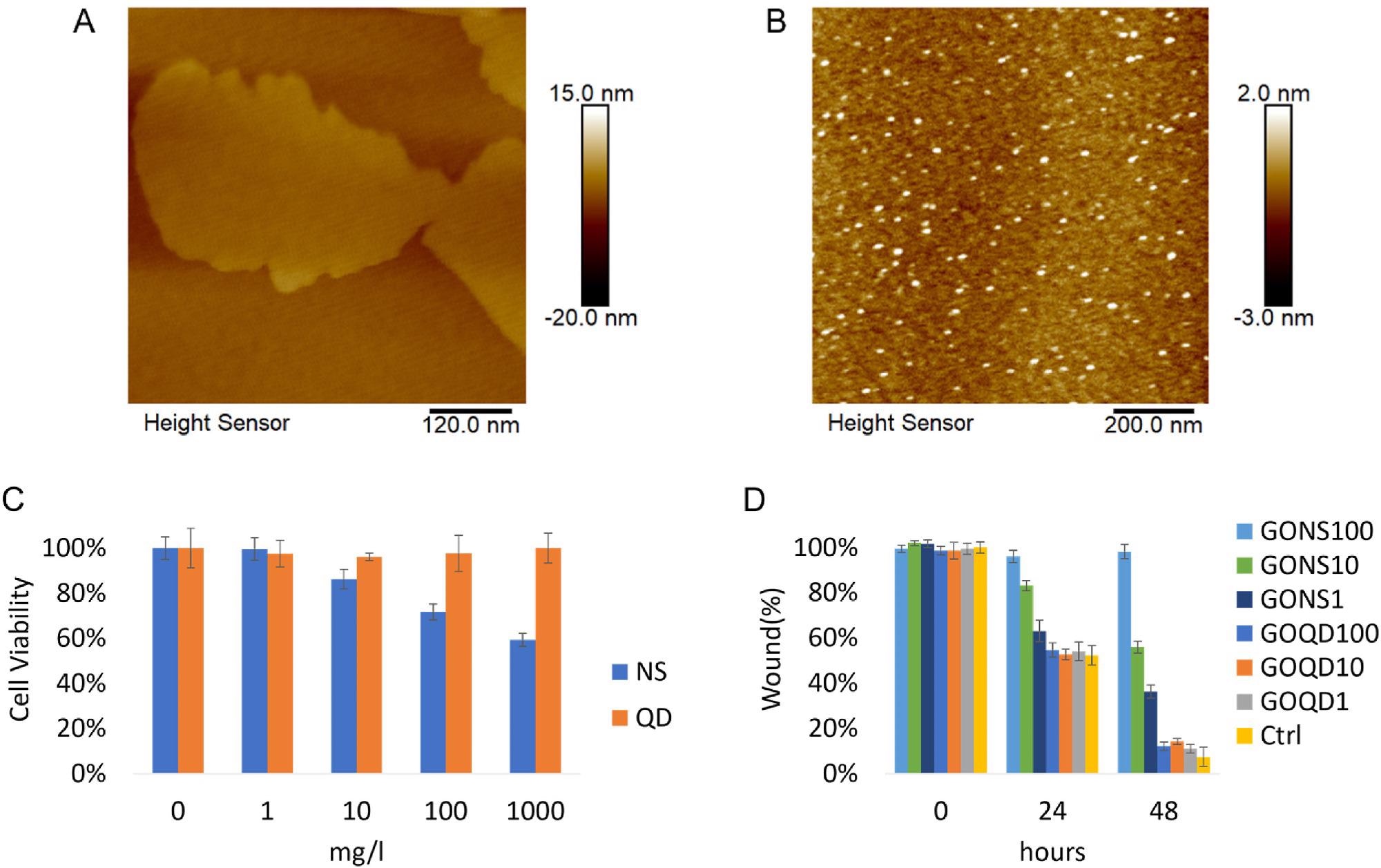
Figure 1. A) Representative AFM image of GO nanosheets. B) Representative AFM image of GO quantum dots. C) Cell viability of LX-2 incubated with GO nanosheets and quantum dots were assessed with CCK-8 assay after 24 h incubation. (*p
