The technique combines Fourier-plane polarimetry and multipole retrieval with laser scanning microscopy to reconstruct sub-diffraction-limit nanoparticle ensembles. It builds on the capabilities of laser scanning microscopy, in which a light beam scans across a sample and measures the light that is transmitted back.
In addition to measuring the brightness of the light that interacts with the sample, the newly introduced technique measures the parameters encoded in the light field.
Beyond brightness alone, the phase, polarization, and scattering angle of the light also provide information about a sample. To capture the information stored in these characteristics, the researchers examined the spatial resolution of the light’s intensity and polarization.
“The phase and polarization of light, together with its intensity, vary spatially in a way that incorporates fine details about the sample with which it interacts — much like the shadow of an object tells us something about the shape of the object itself,” said professor Peter Banzer, who led the research. “However, much of this information is ignored if only the overall light power is measured after the interaction.”
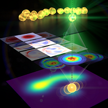
The team validated the experimental results by comparing them to benchmark scanning electron microscopy images.
“Although the particles and their distances were much smaller than the resolution limit of many microscopes, our method was able to resolve them,” Banzer said. “In addition, and even more importantly, the algorithm was able to provide other parameters about the sample, such as the precise size and position of the particles.”
Most microscopy techniques that can resolve images beyond the diffraction limit, such as superresolution microscopy, require the sample to be modified with fluorescent labels. Superresolution techniques that do not rely on any form of sample modification have a limited capability to enhance resolution.
“Our novel approach to laser-scanning microscopy could close the gap between conventional microscopes with limited resolution and superresolution techniques that require modification of the specimen under study,” Banzer said. “In comparison to superresolution techniques based on a similar scanning approach, our method is fully noninvasive, meaning it doesn’t require any fluorescent molecules to be injected into a specimen before imaging.”
Now, the researchers are adapting the method so that it can be used with more complex samples. They believe the functionality of the method could be expanded by tailoring the structure of the light that interacts with the sample and incorporating artificial intelligence-based approaches into the image processing steps.
The team is also contributing to the development of a camera that will be capable of resolving polarization and phase information, in addition to intensity. The development of this next-generation detection device is part of a European project called SuperPixels.
Measuring the distribution of nanoparticles is a common task in science, and the technique could be used in many scientific disciplines to reconstruct complicated nanostructures and arrangements of structures. “Our approach could help extend the microscopy toolbox used to study nanostructures in a variety of samples,” Banzer said. “Our study is another demonstration of the pivotal role that the structure of light can play in the field of optics and light-based technologies.”
The research was published in Optica (www.doi.org/10.1364/OPTICA.450712).
