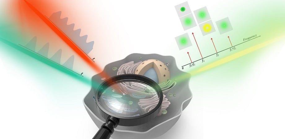
STED microscopy was developed in 1994, demonstrated in 1999, and earned its developer, Stefan Hell, the Nobel Prize in chemistry in 2014. The practical use of STED microscopy, which is a superresolution technique to image beyond the capabilities of nanoscopy or the generally accepted optical limit of 200 to 300 nm, involves undesirable background noise that affects spatial resolution and image quality. In general, it comes from two signal sources: fluorescence generated by reexcitation caused by ultrahigh light doses from the depletion beam, and residual fluorescence due to insufficient depletion of the inhibition beam.
Though background removal approaches have been developed, limitations of their implementation range from image distortion to prolonged acquisition times, and/or the introduction of shot noise.
Existing methods for background removal approaches can be divided into the categories of time-domain, space-domain, and phasor-domain.
Since it avoids the re-excitation caused by the depletion beam, a depletion laser with a wavelength closer to the peak of the fluorescence emission spectrum of the sample can be selected, which reduces the required depletion intensity.
The current version of dmdSTED performs with spatial resolution of ?/8, which is higher resolution than that of the phasor-domain methods, which are prone to shot noise. Theoretically, the researchers said, the approach can help to avoid potential signal loss by time-domain approaches, such as time-gating.
In addition, dmdSTED is compatible with either pulsed or continuous-wave scenarios, and hardware for time-correlated single-photon counting is not required for use. Compared with space-domain methods, the time resolution of dmdSTED is not confined. As a result, dmdSTED is advantageous in the acquisition of comprehensively fine microscopy images, in spatial resolution, signal-to-noise ratio, and time resolution.
According to Xu Liu, director of the State Key Laboratory of Modern Optical Instrumentation and senior author of the study, the frequency domain method has the potential to be integrated into other dual-beam point-scanning techniques. These include excited state saturation microscopy, charge state depletion microscopy, and ground state depletion microscopy.
Further, Liu said, the method can accept more types of samples with spectral characteristics that differ from the fluorescent dyes that are commonly used in STED imaging. These include some quantum dots that may demonstrate wider excitation spectra.
The research was published in Advanced Photonics (www.doi.org/10.1117/1.AP.4.4.046001).
