| Date | 2nd, Jan 2023 |
|---|
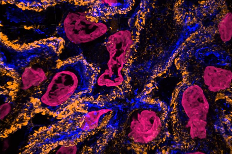
Expansion microscopy (ExM) provides unprecedented views of cell interiors. The emerging super-resolution imaging technique relies on physical — rather than optical — magnification. Advancements by Carnegie Mellon University’s Zhao Biophotonics Lab increases the expansion rate and allows many types of tissues to be viewed in 3D. Credit: Carnegie Mellon University
Unprecedented views of the interior of cells and other nanoscale structures are now possible thanks to innovations in expansion microscopy. The advancements could help provide future insight into neuroscience, pathology, and many other biological and medical fields.
In the paper “Magnify is a universal molecular anchoring strategy for expansion microscopy,” published today (January 2, 2023) in the journal Nature Biotechnology, collaborators from Carnegie Mellon University, the University of Pittsburgh, and Brown University describe new protocols for dubbed Magnify.
“Magnify can be a potent and accessible tool for the biotechnology community,” said Yongxin (Leon) Zhao, the Eberly Family Career Development Associate Professor of Biological Sciences.
Zhao’s Biophotonics Lab is a leader in the field of enabling super-resolution imaging of biological samples through physically expanding samples in a process known as expansion microscopy. Through the process, samples are embedded in a swellable hydrogel that homogenously expands to increase the distance between molecules allowing them to be observed in greater resolution. This allows nanoscale biological structures that previously only could be viewed using expensive high-resolution imaging techniques to be seen with standard microscopy tools.
